Download
1 / 55
550 likes | 675 Views
BIOLOGY OF HUMAN AGING. CHAPTER 8 The Special Senses. Eye and Associated Structures. 70% of all sensory receptors are in the eye Most of the eye is protected by a cushion of fat and the bony orbit

E N D
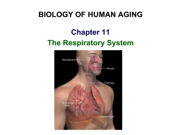
BIOLOGY OF HUMAN AGING CHAPTER 8 The Special Senses
Eye and Associated Structures • 70% of all sensory receptors are in the eye • Most of the eye is protected by a cushion of fat and the bony orbit • Accessory structures include eyebrows, eyelids, conjunctiva, lacrimal apparatus, and extrinsic eye muscles
Conjunctiva • Transparent membrane that: • Covers the whites of the eyes as the ocular conjunctiva • Lubricates and protects the eye
Lacrimal Apparatus • Consists of the lacrimal gland and associated ducts • Lacrimal glands secrete tears • Tears • Contain mucus & antibodies • Drain into the nasolacrimal duct
Lacrimal Apparatus Figure 15.6
Extrinsic Eye Muscles Figure 15.7a, b
Structure of the Eyeball • A slightly irregular hollow sphere with anterior and posterior poles • The wall is composed of three tunics – fibrous, vascular, and sensory • The internal cavity is filled with fluids called humors
Structure of the Eyeball Figure 15.8a
Fibrous Tunic • Forms the outermost coat of the eye and is composed of: • Opaque sclera (posteriorly) • Clear cornea (anteriorly) • The sclera protects the eye and anchors extrinsic muscles • The cornea lets light enter the eye
Vascular Tunic: Choroid Region • Has three regions: choroid, ciliary body, and iris • Choroid region • A dark brown membrane • Supplies blood to all eye tunics
Vascular Tunic: Ciliary Body • A thickened ring of tissue surrounding the lens • Composed of smooth muscle bundles (ciliary muscles) • Anchors the suspensory ligament that holds the lens in place
Vascular Tunic: Iris • The colored part of the eye • Pupil – central opening of the iris • Regulates the amount of light entering the eye during: • Close vision and bright light – pupils constrict • Distant vision and dim light – pupils dilate
Sensory Tunic: Retina • A delicate two-layered membrane • Pigmented layer – the outer layer that absorbs light and prevents its scattering • Neural layer, which contains: • Photoreceptors that transduce light energy
Sensory Tunic: Retina Figure 15.10a
The Retina: the Optic Disc The optic disc: • Is the site where the optic nerve leaves the eye • Lacks photoreceptors (the blind spot)
The Retina: Optic Disc Figure 15.10b
The Retina: Photoreceptors • Rods: • Respond to dim light • Are used for peripheral vision • Cones: • Respond to bright light • Have high-acuity color vision • Are found in the macula lutea • Are concentrated in the fovea centralis
Inner Chambers and Fluids • The lens separates the internal eye into anterior and posterior segments • The posterior segment is filled with a clear gel called vitreous humor that: • Transmits light • Supports the posterior surface of the lens • Holds the neural retina firmly against the pigmented layer
Anterior Segment • Composed of two chambers • Anterior – between the cornea and the iris • Posterior – between the iris and the lens • Aqueous humor • A plasma like fluid that fills the anterior segment • Supports, nourishes, and removes wastes
Anterior Segment Figure 15.12
Lens • A biconvex, transparent, flexible, avascular structure that: • Allows precise focusing of light onto the retina • Lens epithelium – anterior cells that differentiate into lens fibers • With age, the lens becomes more compact and dense and loses its elasticity
Refraction and Lenses • When light passes from one transparent medium to another its speed changes and it refracts (bends) • Light passing through a convex lens (as in the eye) is bent so that the rays converge to a focal point
Problems of Refraction • Emmetropic eye – normal eye with light focused properly • (nearsighted) – the focal point is in front of the retina • Corrected with a concave lens • (farsighted) – the focal point is behind the retina • Corrected with a convex lens
Problems of Refraction Figure 15.18
Adaptation • Adaptation to bright light (going from dark to light) involves: • Dramatic decreases in retinal sensitivity – rod function is lost • Switching from the rod to the cone system – visual acuity is gained • Adaptation to dark is the reverse • Cones stop functioning in low light
Age-related changes Visual problems Slight shrinkage and degeneration of some cells. Increase in the amount of connective tissues. Reduced blood supply. Loss of fat and elastic tissues thinning of the skin in the eyelids (bags). Dry cornea irritation and inflammation Production of tears by the lacrimal gland is reduced with aging. Visual problems Increase in amount of connective tissues Degeneration of some cells Conjuctiva become thinner and more fragile Astigmatism Deposition of calcium and cholesterol salts senile ring Ciliary body produces less aqueous humor affecting nourishment and cleansing of the lens atrophy of muscle cells General decrease of photoreceptors Photopigement (rhodopsin) decreases with aging Lipofuscins accumulates in the retina Light flashes
Inclusion bodies floaters may increase with aging Iris become hardened with aging and atrophy of the dilator muscles senile miosis Poor drainage of aqueous humor Lens become yellow and less transparent with age Lens change shape Atherosclerosis of choroid and blood vessels of the retina Adaptation takes longer Less light reaching the retina sensitivity to light decreases with aging Slower scanning process in the brain takes longer for older persons to identify objects
Age-related Dysfunction Presbyopia Blindness Glaucoma Diabetic retinopathy Cataracts Macular degeneration Detached retina
The Ear: Hearing and Balance • The three parts of the ear are the inner, outer, and middle ear • The outer and middle ear are involved with hearing • The inner ear functions in both hearing and equilibrium
The Ear: Hearing and Balance Figure 15.25a
Outer Ear • The auricle (pinna) • External auditory meatus • Short, curved tube Tympanic membrane (eardrum) • Thin connective tissue membrane that vibrates in response to sound • Transfers sound energy to the middle ear ossicles • Boundary between outer and middle ears
Middle Ear (Tympanic Cavity) • A small, air-filled, mucosa-lined cavity • Flanked laterally by the eardrum • Flanked medially by the oval and round windows • Auditory tube – connects the middle ear to the nasopharynx • Equalizes pressure in the middle ear cavity with the external air pressure
Middle Ear (Tympanic Cavity) Figure 15.25b
Ear Ossicles • The tympanic cavity contains three small bones • Transmit vibratory motion of the eardrum to the oval window
Inner Ear • Bony labyrinth • Tortuous channels worming their way through the temporal bone • Contains the vestibule, the cochlea, and the semicircular canals • Filled with perilymph
Inner Ear Figure 15.27
The Vestibule • The central egg-shaped cavity of the bony labyrinth • Suspended in its perilymph are two sacs: the saccule and utricle • The saccule extends into the cochlea • The utricle extends into the semicircular canals • These sacs: • House equilibrium receptors called maculae • Respond to gravity and changes in the position of the head
The Semicircular Canals • It houses equilibrium receptors • These receptors respond to angular movements of the head
The Semicircular Canals Figure 15.27
The Cochlea • A spiral, conical, bony chamber that: • Extends from the anterior vestibule • Contains the cochlear duct • Contains the organ of Corti (hearing receptor)
Sound and Mechanisms of Hearing • Sound vibrations beat against the eardrum • The eardrum pushes against the ossicles, which presses fluid in the inner ear against the oval and round windows • This movement sets up shearing forces that pull on hair cells • Moving hair cells stimulates the cochlear nerve that sends impulses to the brain via vestibulocochlear nerve
Resonance of the Basilar Membrane Figure 15.32
Mechanisms of Equilibrium and Orientation • Vestibular apparatus – equilibrium receptors in the semicircular canals and vestibule • Maintains our orientation and balance in space • Vestibular receptors monitor static equilibrium • Semicircular canal receptors monitor dynamic equilibrium
Age-related changes • Auricle become long, wide, and lose flexibility • Auditory meatus becomes wider and stiffer • Excessive earwax • Joints between ossicles may ossify less freely movable problem in vibration • Degeneration of spiral organ cells • Thickening of the walls of capillaries supplying the cochleadecrease in nutrients • Decrease in the number of nerve fibers problems in balance and coordination
Age-related Dysfunction Presbycusis Tinnitus Deafness Dizziness and Verdigo
Taste& Smell The Special Senses
Chemical Senses • Chemical senses – gustation (taste) and olfaction (smell) • Their chemoreceptors respond to chemicals in aqueous solution • Taste – to substances dissolved in saliva • Smell – to substances dissolved in fluids of the nasal membranes
Taste Buds • Most of the 10,000 or so taste buds are found on the tongue • Microvilli
Taste Buds Figure 15.1