
The Role of First Transcribed Nucleoside in Human Cap-Binding Complex Interaction
Understanding the importance of the first transcribed nucleoside in human nuclear cap-binding complex interaction can provide insights into mRNA processing and translation initiation. This study explores the energetic description and binding affinity of different cap analogues to the CBC. Results show a preference for dinucleotide cap analogues over mononucleotides due to stronger binding energies. The study highlights the significance of nucleoside composition in determining the stability of the cap-CBC complex.

The Role of First Transcribed Nucleoside in Human Cap-Binding Complex Interaction
E N D
Presentation Transcript
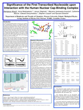
Significance of the First Transcribed Nucleoside upon Interaction with the Human Nuclear Cap-Binding Complex Remigiusz Worch1, Anna Niedzwiecka1,3, Janusz Stepinski 1, Marzena Jankowska-Anyszka2, Catherine Mazza4, Edward Darzynkiewicz 1, Stephen Cusack4 and Ryszard Stolarski1 1Department of Biophysics and 2Faculty of Chemistry, Warsaw University, Poland, 3Biological Physics Group, Institute of Physics PAS, Warsaw, 4EMBL, Grenoble, France Introduction All nuclear transcribed mRNAs possess a 5'-terminal "cap", m7GpppN, in which 7-methylguanosine is linked by a 5’-5’-triphosphate bridge to the first transcribed nucleoside. Recognition of the mRNA 5’ cap structure by the nuclear protein Cap Binding Complex (CBC) plays a key role in pre-mRNA splicing, polyadenylation of the 3' terminus, U snRNA transport, nonsense-mediated decay and translation initiation. The CBC is a heterodimer consisting of two subunits: CBP20 and CBP80. The former directly interacts with the cap structure while the latter is essential for the active complex formation. The previously known crystal structures of the apo-CBC (1) and the CBC in the complex with a cap analogue, m7GpppG (2,3), showed large conformational changes of the N- and C- terminal parts of CBP20 upon mRNA 5’cap binding. The m7G moiety of 5’ mRNA cap interacts with the CBC by sanwich stacking with Tyr20 and Tyr 43 enhanced by numerous hydrogen bonds, and the first transcribed nucleoside stacks with Tyr 138 (Scheme 1). While the 3D structure of the protein was solved, the consistent energetic description of the cap-CBC interaction was lacking. To address the mechanistic bases of complex formation and stability, solution studies were necessary. Because the CBC has many fluorescent residues and cap binding results in quenching of the intrinsic protein fluorescence due to conformational changes, a method of time-synchronized titration (4) was used in order to analyze the molecular basis of the cap affinity for the CBC. By means of this method, including the activity of the non-enzymatic protein and emission of all species explicitly into the analysis, we have determined equilibrium association constants (Kas) for a series of mono- and dinucleotide cap analogues (Scheme 2) and with a tetranucleotide mRNA fragment, m7GpppAm2’pUm2’pAm2’. The Kas values can be interpreted in terms of Gibbs free energy of binding (G ). The role of stacking between Tyr138 of the CBC and the base moiety of the first transcribed nucleotide was examined by determining Kas for a group of cap analogues with the Y138A mutant. Scheme 2 All used cap analogues were triphosphates (n=3) m7GTP R1=CH3 R2=R3=H m22,7GTP R1=CH3 R2=CH3, R3=H m32,2,7GTP R1=CH3 R2, R3= CH3 et7GTP R1=CH2CH3 R2=CH3, R3=H m7GpppG R1=CH3 R2=R3=H R4=OH B=G m7GpppGm2' R1=CH3 R2=R3=H R4=OCH3 B=G m7GpppA R1=CH3 R2=R3=H R4=OH B=A m7Gpppm6A R1=CH3 R2=R3=H R4=OH B=m6A m7GpppAm2' R1=CH3 R2=R3=H R4=OCH3 B=A m7Gpppm7G R1=CH3 R2=R3=H R4=OH B=m7G et7GpppG R1= CH2CH3 R2=R3=H R4=OH B=G m7GpppU R1=CH3 R2=R3=H R4=OH B=U m7GpppC R1=CH3 R2=R3=H R4=OH B=C m22,7GpppG R1=CH3 R2=CH3, R3=H R4=OH B=G m32,2,7GpppGR1=CH3 R2,R3=H R4=OH B=G A tetranucleotide mRNA fragment m7GpppAm2'pUm2'pAm2 consists of m7GpppAm2' with linked U andA. Scheme 1 Results & Discussion CBC significantly prefers to bind the monomethylated dinucleotide cap analogs comparing with the mononucleotides, except for the compounds hipermethylated at the N2 amino group of the 7-methylguanosine moieties. The average binding energy of the dinucleotides is by ~1 kcal/mol greater (more negative) than that for m7GTP, which is accompanied by a drop of one order of magnitude in terms of the association constant values (Table, Figure2). This preference agrees with former results of in vitro and in vivo biological experiments (5). The titration experiments with a tetranucleotide mRNA fragment, m7GpppAm2’pUm2’pAm2’ yielded slightly smaller binding energy than that for the corresponding control dinucleotide, m7GpppAm2’. This suggests that only two first nucleotides at the mRNA 5’ terminus are responsible for the tight, specific interaction with the CBC. However, further nucleotides may be involved in nonspecific contacts with the protein. The differences of G should be ascribed to the entropic costs of ordering of the longer oligonucleotide chain upon the binding. The purine base in the first transcribed nucleoside is preferred. The average energy of binding for the dinucleotides with pyrimidine (C or U) as the second base is by ~0.4 kcal/mol less than the average binding energy for the purine (A or G) dinucleotides, and their Kas values are ~2-fold lower, respectively. m7GpppG is bound stronger than m7GpppA unless adenosine is additionally methylated at the amino group or at 2'-hydroxyl. The Y138 mutant binds m7GpppG with Kas ~4-fold lower than the wild type CBC leading to a decrease of G by 0.80 ± 0.36 kcal/mol, whereas for m7GTP the change is negligible. A difference in a mode of m7GpppG binding is observed: while for the wild type CBC the fitting residuals have non-random distribution resulting from systematic deviations (Figure 1a)), in case of the Y138A mutant the deviations are diminished (Figure 2b)). For the wild type CBC, these deviations appeared for the dinucleotides containing pyrimidine as the second base and not for the purine dinucleotides. The modes of binding of the hipermethylated mono- and dinucleotide cap analogs by CBC are distinctly different. Whereas introduction of either one or two methyl groups at the amino group of 7-methylguanosine results in a dramatic diminution of Kasby ~100-fold (decrease of G by ~2.5 kcal/mol) for the dinucleotides, the Kas values for the corresponding mononucleotides surprisingly change only less than 2-fold. The total fluorescence quenching pattern also reveals the differences. It is observed only a few percent of quenching for m32,2,7GTP while it is up to 12 % for m32,2,7GpppG.This surprising discrepancy is the same for the Y138A mutant (Table, Figure 2) whichpoints to the steric hindrancecaused already by the first methylation at the m7G amino group. Thisprevents the hipermathylated cap from deep penetration into the binding pocket. Figure 1. Table. Equilibrium association constants (Kas) and binding Gibbs free energie (DG0) for various cap analogues-CBC (wilde type) and cap-Y138A mutant complexes at 20C. Figure 2. Graphical presentation of the Gibbs free energies of association of the cap analogs with a) wild type CBC, and b) Y138A mutant. Conclusions The presented results together with the structural view known from crystallography make the first step to build molecular mechanism of specific recognition and binding between CBC and mRNA 5’ cap. The enhanced affinity forthe dinucleotide cap analogues, especially these containing pyrimidine as the second base, as well asdifferent modes of binding of mono- and dinucleotides point at the significance of the first transcribed mRNA nucleoside for the CBC recognition. The resultsindicate the possibility that the two-partite cap-binding center of the CBC can bind the first and the second nucleosides belonging to cap with some, still unknown, level of cooperativity. References 1. Mazza, C., Ohno, M., Segref, A., Mattaj, I. W., & Cusack, S. (2001) Mollecular Cell 8, 383–396. 2. Mazza, C., Segref, A., Mattaj, I. W., & Cusack, S. (2002) The EMBO Journal 21, 5548–5557. 3. Calero, G.,Wilson, K., Ly, T., Rios-Steiner, J. R., Clardy, J., & Cerione, R. A. (2002) Nature Struct. Biol. 9, 912–917. 4. Niedzwiecka, A., Marcotrigiano, J, Stepinski, J., Jankowska-Anyszka, M., Wyslouch-Cieszynska, A., Dadlez, M., Gingras, A.-C., Mak, P., Darzynkiewicz, E., Sonenberg, N., Burley, S. K., and Stolarski, R. (2002) J. Mol. Biol. 319, 615–635. 5. Izaurralde, E., Lewis, J., McGuigan., Jankowska, M., Darzynkiewicz, E., and Mattaj, I.W. (1995) Cell78, 657–668. Acknowledgements This work was supported by the State Committee for Scientific Research KBN 3 P04A 021 25, PBZ/KBN/059/T09/2001, BST 833/BF. We are indebted to Teija Nittymaki and Harri Lönnberg for cooperation in preparing the tetranucleotide.


