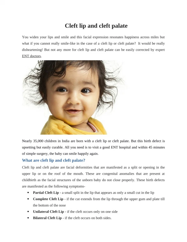
Alveolar cleft repair
520 likes | 1.92k Views
Brett A. Ueeck DMD, MD, FACS. Alveolar cleft repair. History of Alveolar Repair. Management with bone grafting first described in the early 20 th century Lexer 1908, Drachter 1914 50 years passed without any progress Mid 1950’s increase in primary bone grafting

Alveolar cleft repair
E N D
Presentation Transcript
Brett A. UeeckDMD, MD, FACS Alveolar cleft repair
History of Alveolar Repair • Management with bone grafting first described in the early 20th century • Lexer 1908, Drachter 1914 • 50 years passed without any progress • Mid 1950’s increase in primary bone grafting • 1960’s: Increasing evidence primary bone grafting had negative impact on growth • 1970’s: Secondary bone grafting introduced by Boyne/Sands and becoming treatment of choice • 1980-90’s: Continued research in donor sites, alveolar molding, GPP, growth and overall outcomes • Early 21st Century: rhBMP-2, Stem cells, implants
The Alveolar Defect • Skeletal • Collapse and rotation of the maxillary segments • Cleft in alveolus to piriform rim • Soft Tissue • Lack of attached tissue • Absent gingival anatomy • Oral-nasal fistula • Dental • Missing permanent teeth • Malformed teeth • Supernumerary teeth • Impacted teeth
Goals Of Alveolar Repair Success Defined By: 1Grafting must achieve stability of the arch and prevent collapse of the alveolar segments 2Grafting must preserve the health of the dentition and maintain bony support of teeth adjacent to the cleft 3Grafting must restore continuity not only of the alveolus but also the anterior hard palate and the maxilla at the piriform rim (support for alar base) 4Grafting must support the soft tissue closure of the oronasalfistula 5Grafting must have adequate volume of bone matrix for erupting teeth in the line of the cleft, and for orthodontic movement of the involved teeth into appropriate “nontorqued” position in the dental arch 6 Grafting must allow for the successful placement of dental implant(s) Tai et al. J Oral MaxillofacSurg58:1241-1249, 2000
Timing of Repair • Early • Gingivo-perioplasty (with primary lip and/or palate) • Primary bone grafting (abandoned) • Secondary (Traditional) • Age 7-13 • Autogenous Bone v rhBMP-2 • Tertiary Repair (Late) • After completion of growth • With or without maxillary orthognathic surgery • ICBG with and without rhBMP2 • Completion of Repair • Bone and soft tissue repair • Dental implant placement
Early Repair • Gingivoperioplasty • Originally described in 1965 by Skoog • Advanced by Millard, Lathum, Grayson • Closes the alveolus with the lip and/or palate • Combined with dentofacial orthopedic techniques to reduce the cleft width • NAM, Lathum, Tapping, Lip Adhesion Skoog, T. The use of periosteal flaps in the repair of clefts of the primary palate. Cleft Palate Craniofac. J. 2: 232, 1965.
Gingivoperioplasty • Success • Quantity of bone • Quality of bone • Normal eruption of permanent dentition • Elimination of fistula • Effect on Growth • Dental arch • Maxillary • Facial
Gingivoperioplasty • Success on Bone Formation – Unilateral Clefts • 60% of GPP cases did not need secondary grafting • 73% of GPP cases did not need secondary grafting • Higher incident of secondary grafting needed in the bilateral cleft cases. • 66% need secondary grafts SantiogoCleft Palate–Craniofacial Journal, January 1998, Vol. 35 No. 1 Sato, Plast. Reconstr. Surg. 121: 1356, 2008 SantiogoCleft Palate–Craniofacial Journal, January 1998, Vol. 35 No. 1
Gingivoperioplasty • Success on Fistula Outcome • Overall GPP nearly eliminates fistula • GPP eliminates fistula in every case in one study Sato, Plast. Reconstr. Surg. 121: 1356, 2008
Gingivoperioplasty • Effect on Growth • Early GPP (Skoog) • Conclusive: Negative Impact • Millard and Lathum • Somewhat inconclusive • Patient follow-up, number of patients, etc • Grayson • No negative growth outcomes • Matic • Potential for growth attenuation
Gingivoperioplasty • Effect on the Dentition – Quite Variable Negative Outcomes Positive Outcomes • Missing primary teeth and alteration in eruption • Missing permanent central incisors and higher rate of missing lateral incisors • Malformed teeth • Normalized eruption of primary teeth • No difference in number of missing teeth • Better periodontal health and support for permanent teeth adjacent to the cleft • Stabilized arches
Gingivoperioplasty • Overall • Holds promise for superior alveolar repair • Requires a dedicatedandwell trained team for best results • Surgeon dependent – technique sensitive
Secondary Bone Grafting • Current preferred method of repair • Timing • Early • Age 7-9: At the time of central incisor eruption • Late • Age 9-11: Canine root 2/3rds formed • Autogenous Donor Sites • Iliac Crest » Rib • Cranium » Tibia • Mandible • rhBMP-2
Secondary Bone Grafting • Donor Sites – Autogenous Bone • Iliac Crest Bone Graft • Boyne 1972 – first introduction of secondary graft • Considered the “Gold Standard” • Mandibular Bone Graft • Bosker 1980 • Cranial Bone Graft • Tessier 1982 • J.D. Smith 1973
Difference in Graft Donor Sites ICBG Mandible Cranial • Visible scar • Less pain • No gait issues • Single surgical field • Reliable quantity • Reliable quality • Successful • Visible scar • Pain • Gait disturbance • Second surgical field • Reliable quantity • Reliable quality • Reliable success • No visible scar • Pain • No gait issues • Single surgical field • Quantity? • Reliable quality • Reliable success
Secondary Bone Grafting Considerations… • Impact on Growth • Timing of the graft important • Operative technique? • Selection criteria? • Significant growth retardation – need orthognathic surgery – wait to graft? • Absence of lateral incisor/impacted canine – wait to graft? • Large, unfavorable clefts – 2 stage repair? Single stage with tongue flap?
Secondary Bone Grafting • Further Topics of Consideration • Flap design – degree of periosteal elevation • BCLCP – stage repair? • Splinting post repair • Dressing • PO diet • Antibiotics
Secondary Bone Grafting Facial Growth Outcomes in the Grafted Patient • Ross (1987) – multicenter comparison of UCLCP grafted patients age 4-10 • Marked deficiency in anterior upper-facial height compared to non-grafted patients at age 15 • Grafted group age 9-12 showed no difference in non-grafted group. • Confounded study • Brättstrometal (1992) – comparison of 85 UCLCP pts with primary graft, secondary graft, and no graft • Primary – poor growth • Secondary – after incisors before canine had better growth • Non-grafted patients had the best growth
Secondary Bone Grafting Facial Growth Outcomes in the Grafted Patient • Semb (1988) – National Center of Logopedics, Oslo, Norway • no difference in growth among UCLCP children grafted between the ages of 8 and 12 compared to non-grafted patients
Effect of Alveolar Bone Grafting in the Mixed Dentition on Maxillary Growth in Complete Unilateral Cleft Lip and Palate Patients • John Daskalogiannakis, D.D.S., M.Sc.R. Bruce Ross, D.D.S., M.Sc. • The Cleft Palate-Craniofacial Journal: Vol. 34, No. 5, pp. 455–458. 1997
Grafted group: 8.4-12.9yrs (10.3) • Lateral cephs taken pre-grafting and 5-6years post grafting • John Daskalogiannakis, D.D.S., M.Sc.R. Bruce Ross, D.D.S., M.Sc. • The Cleft Palate-Craniofacial Journal: Vol. 34, No. 5, pp. 455–458. 1997
Conclusions • Secondary bone grafting in the UCLCP patient in the age range 8-13yrs of age does not seem to negatively impact the vertical or AP maxillary growth and development. • John Daskalogiannakis, D.D.S., M.Sc.R. Bruce Ross, D.D.S., M.Sc. • The Cleft Palate-Craniofacial Journal: Vol. 34, No. 5, pp. 455–458. 1997
Secondary Bone Grafting • rhBMP-2 • Recombinant Human Bone Morphogenic Protein 2 • First discovered in 1965 by Dr. Marshall Urist • Now available for clinical use via Medtronic as Infuse® Bone Graft
rhBMP-2 • Growth Factor • From the superfamily TGF-B • Several different BMPs induce bone formation • 2, 4, 7 • BMP-2 acts early in the cascade of events and directly triggers mesenchymal cells to become osteoblasts
rhBMP-2 • The bovine ACS (absorbable collagen sponge) is important for the clinical effects of BMP-2. Binds 95% in 15 minutes. Elutes the BMP-2 for 8-10 days. Fully resorbed in 4-10weeks. Release kinetics studies show that the ACS releases bound rhBMP-2 in a time period optimal for bone formation
rhBMP-2 Important steps to success • Create appropriate space for rhBMP 2 + ACS • The ACS cannot maintain the space alone • Must ensure access to cells • Periosteum • Adjacent bleeding bone • Proper preparation of the material
rhBMP-2 • The Science in Alveolar Clefts
rhBMP-2 • ICBG: 78% bone fill • rhBMP-2: 72% bone fill Average Bone Fill
rhBMP-2 • Benefits • Eliminates need for second (donor) surgical site • Eliminates donor site morbidity • Decreased OR time • Decreased hospital LOS • Decreased health care cost
rhBMP-2 Effective Alternative to ICBG • Conclusions are the same as JOMS 2007 • Eliminates need for second (donor) surgical site • Eliminates donor site morbidity • Decrease in surgical site complications • Decreased OR time • Decreased hospital LOS • Decreased health care cost
Retrospective study • 23 implants placed in 21 patients • 5 patients not eligible for inclusion • 16 patients (8 male, 8 female) (Oral Surg Oral MedOralPathol Oral RadiolEndod 2008;105:297-302)
Secondary Soft Tissue Management • Keratinized grafting in cleft site • Improve peri-implant tissue health • Scar revision • Improve vestibule characteristics • Connective tissue grafting • Improve prosthetic appearance