Download
1 / 60
640 likes | 861 Views
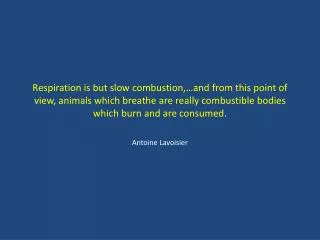
Respiration is but slow combustion,…and from this point of view, animals which breathe are really combustible bodies which burn and are consumed. Antoine Lavoisier. Acute Respiratory Failure. Overview. Physiology/Pathophysiology Types of respiratory failure Diagnosis ABG CXR Treatment

E N D
Respiration is but slow combustion,…and from this point of view, animals which breathe are really combustible bodies which burn and are consumed. Antoine Lavoisier
Overview • Physiology/Pathophysiology • Types of respiratory failure • Diagnosis • ABG • CXR • Treatment • Oxygen supplementation • Non-Invasive Ventilatory support
Physiology • Many components constitute an intact respiratory system: • Central Nervous System • Peripheral Nervous System • Respiratory muscles • Chest Wall • Airways • Alveoli • Cellular Activity • Electron Transport System
Pathophysiology • Several types of respiratory failure • Acute vs. Chronic • Hypoxemic (type I) vs. --Hypercapneic (type II) • Many clinical situations have overlap of these broad categorizations; ‘combined’. • type III
Hypoxemic Respiratory FailureType I • Defined as an inadequate PaO2 despite high levels of supplemental oxygen. • Chronicity may sometimes be appreciated by accompanying polycythemia or cor pulmonale and absence of acute mental status changes.
Type I Failure • ARDS • Asthma • Atelectasis • Cardiogenic Pulmonary Edema • COPD • Interstitial Fibrosis • Pneumonia • Pulmonary embolism
Mechanisms of Hypoxemia • 5 major causes of hypoxemia • Nl A-a gradient • Alveolar Hypoventilation • Altitude • Widened A-a gradient • Ventilation-Perfusion Mismatch • Shunt • Diffusion Limitation
Alveolar Hypoventilation • Defined by decreased respiratory drive, these disorders are exemplified by CNS derangements: • Drug Overdose (narcotics, sedatives,etc.) • Structural • Mass effects • CVA’s (medullary control center) • Meningoencephalitis • Metabolic- myxedema, uremia, met. Alkalosis • Obesity-Hypoventilation syndrome • Unknown mechanism
Shunt • Deoxygenated mixed venous blood either bypasses ventilated alveoli or supplies poorly ventilated alveoli, resulting in hypoxemia relative to the shunt fraction. • Intra-cardiac • Extra-cardiac • Intra-pulmonary • Extra-pulmonary
V/Q Mismatch • Areas of disproportionately low ventilation relative to perfusion contribute to hypoxemia.
Diffusion Limitation • Impaired, or increased, pathway from alveolar space to capillary membrane resulting in decreased oxygen transport across the alveolar membrane.
Hypercapneic Respiratory FailureType II • Defined as an arterial PCO2 >45 mmHg. • Acute Hypercapneic respiratory failure includes accompanying acidemia (pH<7.3).
Ventilation Physiology VA= K . VCO2 / PaCO2 • VA= minute alveolar ventilation • K = a constant • VCO2= the rate of CO2 production
Ventilation Physiology VA is dictated by: • VE= minute ventilation; and • VD/VT= the dead space to tidal volume ratio SO,
Ventilation Physiology VE= K . (VO2 . RQ) PaCO2 / (1- VD/VT) • VO2= rate of O2 consumption • RQ= the respiratory quotient • VD= dead space volume • VT= tidal volume
Ventilation:Supply vs. Demand • Ventilatory Supply is the maximal spontaneous ventilation that can be sustained without development of respiratory muscle fatigue. • Ventilatory Demand is the spontaneous minute ventilation which, when constant, results in a stable PaCO2.
Factors that Increase Ventilatory Demand • Increased VD/VT= Asthma, COPD, PE • Increased VO2= Fever, sepsis, trauma, obesity, increased work of breathing • Increased RQ= Overfeeding • Decreased PaCO2= Hypoxemia, sepsis metabolic acidosis, renal or hepatic failure
Physiology • Many components constitute an intact respiratory system: • Central Nervous System • Peripheral Nervous System • Respiratory muscles • Chest Wall • Airways • Alveoli • Cellular Activity • Electron Transport System
Factors that Diminish Ventilatory Supply • Decreased Muscle Strength • Fatigue • Disuse Atrophy • Electrolyte Abnl • “Disadvantaged Diaphragm” • Decreased Motor Neuron Function • Phrenic nerve injury • Decreased NMJ transmission • Disproportionate myoperfusion • High Elasticity • High Resistance • Abnormal Respiratory Mechanics • Airflow Limitation • Loss of Volume • Other restrictive defects
Type III-Combined • Acute Respiratory Distress Syndrome • Asthma • Chronic Obstructive Pulmonary Disease
Diagnosis • Clinical suspicion for any type of respiratory failure may be confirmed by an arterial blood gas (ABG). • Organ-specific, as well as systemic symptoms are assessed to identify likely underlying etiologies.
Physical Examination • Two Pronged Approach • CT Angiogram • BNP Level
Physical Exam • Two-pronged approach: • Search for specific cause of respiratory failure: • Unilateral LE edema • Heart murmurs and gallops • Consolidative Breath sounds • Evaluating Current Respiratory Status • RR • Tidal Volume • Use of Accessory Muscles • Paradoxical Abdominal Movements
Signs and Symptoms • Respiratory • Tachypnea, dyspnea, cyanosis • Cardiovascular • Increased CO, tachycardia, palpitations, arrhythmias, angina, hypotension, diaphoresis, shock • Central Nervous System • Headache, confusion, euphoria, delirium, restlessness, papilledema, seizures, coma • Metabolic • Sodium/Water retention, lactic acidosis
Pulse Oximetry Monitoring • Pulse oximetry is a way of measuring O2 saturation by a noninvasive method which relies on the different absorption characteristics of oxyhemoglobin and deoxy hemoglobin for red (or infrared) light. • The error of pulse oximetry is only around + or - 4% above the saturation of 70%. When used to measure O2 saturation below 70%, the error becomes unacceptably high.
Pulse Oximetry Monitoring • Pulse oximetry can yield falsely elevated O2 saturation in smokers and victims of carbon monoxide poisoning • Pulse oximetry can yield falsely low values in individuals: • given intravenous methylene blue or indocyanine green • who are African-American • with green or blue nail polish • who are in the presence of arc surgical lights, or fluorescent lights
Pulse Oximetry • Rapid, non-invasive mechanism for estimating SaO2. • Useful in initial triage of patients in respiratory distress as well as re-assessing treatments, including oxygen supplementation. • Limitations • No ventilatory data • Ignores Work of Breathing • Unreliable: on steep portion of oxygen-hemoglobin curve, in pts with carboxyhemoglobin or methemoglobin, and in patients with decreased peripheral perfusion
Role for the Arterial Blood Gas • When dealing with “pure” situations, the ABG, and the calculated A-a gradient, can help narrow the differential diagnosis. • Normal A-a gradient (typically <20 mmHg): • Alveolar Hypoventilation • Altitude • Widened A-a gradient (>20 mmHg) • Shunt • V/Q Mismatch • Diffusion Limitation
Hypoxemic Respiratory Failure • A-a gradient • 148 - 1.2(PaCO2) - PaO2 • [PIO2 - PaCO2/R] - PaO2 • Normal (< 20) • Low FIO2 and hypoventilation • Elevated (> 20) • Shunt, ventilation-perfusion inequalities, and diffusion impairment
Spirometry • Useful in trending airflow, particularly in patients with disorders of ventilation. • Neuromuscular Disease • GBS • MG • Toxins • Botulism • Nerve Agents
Hypoxemia with a clear CXR • COPD/Asthma exacerbation • Pulmonary Embolism • Right-to left Shunt • Microatelectasis
Hypoxemia with a White CXR • Pneumonia • ARDS • Embolism • Pulmonary Edema • Interstitial Lung Disease
Diffuse Lung Lesions • Cardiogenic or increased-pressure edema • Left-ventricular (LV) failure • Acute LV ischemia • Accelerated or malignant hypertension • Mitral regurgitation • Volume overload, particularly with coexisting renal and cardiac disease • Increased permeability or low-pressure edema (ARDS) • Sepsis and sepsis syndrome • Mitral stenosis • Acid aspiration • Multiple transfusions for hypovolemic shock • Near-drowning • Pancreatitis • Air or fat emboli • Cardiopulmonary bypass • Pneumonia • Drug reaction or overdose • Inhalation injury • Infusion of biologics (e.g., interleukin 2) • Ischemia-reperfusion (e.g., postthrombectomy, posttransplantation) • Edema of unclear or "mixed" origin • Reexpansion • Neurogenic • Postictal • Tocolysis-associated • Diffuse alveolar hemorrhage • Microscopic angiitis • Collagen vascular diseases • Goodpasture's syndrome • Severe coagulopathy and bone marrow transplantation • Retinoic acid syndrome
Treatment • Triage • Airway Maintenance • Correction of Hypoxemia & Hypercapnia • Identification of Underlying Cause • Management of Underlying Cause
Triage Factors • Acuity of respiratory failure • Severity of: • Hypoxemia • Hypercapnia • Acidemia • Presence of Co-morbid conditions • Rate of Disease Progression • Available Resources
Correction ofHypoxemia & Hypercapnea • Many modalities to improve both hypoxemia and hypercapnea, short of intubation. • Some techniques are mutually exclusive, while others may improve both derangements. • The correction of hypoxemia is paramount as prolonged hypoxemia is the most immediately life-threatening
Response to Supplemental Oxygen • V/Q mismatch is the most common cause of arterial hypoxemia. • The degree of mismatch responds proportionately to the amount of supplemental oxygen delivered. • Hypoxemia resulting from shunt responds less well to O2 administration. • As shunt fractions approach 20%, even FiO2 of 1.0 results in persistent hypoxemia.
COPD and CO2 Retention • These patients are at increased risk for supplemental O2 administration worsening their ventilatory status secondary to: • V/Q mismatch • Haldane effect • Decreased minute ventilation • Loss of CO2 drive
Correction of Hypoxemia • Low-Flow Systems • Nasal Canula • Simple Mask • Reservoir Mask • Partial Rebreather • Non-Rebreather • High-Flow Systems • Venturi Masks • Reservoir Nebulizer Blenders
Nasal Canula • Max FiO2 around 45%. • Flows above 6L/min do not increase oxygen delivery. • Cheap, widely available • (LPM X 4) + 20 = FiO2
Simple Masks • Require flows above 6/l min to avoid CO2 retention. • Cumbersome; interfere with patient activities
Reservoir Masks • Reservoirs typically measure 600-1000cc. • Still require flows greater than 5L/min to flush reservoir of CO2. • Reservoir masks without one way valves are referred to as partial rebreathing masks • Reservoir masks with one-way valves are referred to as nonrebreathing systems.
High-Flow Systems: • For use when specific FiO2 concentrations are required, like: • Hypercapneic COPD exacerbations • High ventilatory demands may outpace low-flow systems.
Venturi Mask • Based on the Venturi modification of the Bernoulli principle. • Can deliver FiO2 between .24 and .50