Download
1 / 43
430 likes | 622 Views
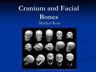
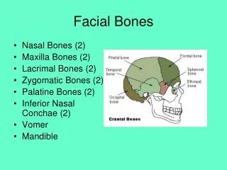
RADT 1522 Orbits, Facial Bones and Nasal Bones. Wynn Harrison, MEd. Facial Anatomy. 14 facial bones (How Many of Each) Maxilla - Vomer Zygomatic - Mandible Palatine Nasal Lacrimal Inferior Nasal Conchae. Mid- saggital view. New Words.

E N D
RADT 1522Orbits, Facial Bones and Nasal Bones Wynn Harrison, MEd
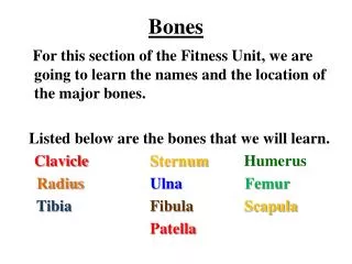
Facial Anatomy • 14 facial bones (How Many of Each) • Maxilla - Vomer • Zygomatic - Mandible • Palatine • Nasal • Lacrimal • Inferior Nasal Conchae
New Words • Blow-Out Fracture – Impact fracture (Trauma) of the orbital floor resulting in orbital intrusion into the maxillary sinus. *** Look at the orbits carefully, since 60 - 70 % of all facial fractures involve the orbit in some way
Le Fort I - tooth bearing portion separated from upper maxilla • Le Fort II - fracture across orbital floor and nasal bridge (pyramidal fracture) • Le Fort III - fracture across frontozygomatic suture line, entire orbit and nasal bridge (craniofacial separation)
LeFort Type I • LeFort Type II • LeFort Type III
Tripod Fracture – A fracture in which the zygoma is separated from its attachment to the maxilla, frontal and temporal bones
Bell’s Palsy - Bell's palsy is a weakness or paralysis of the muscles that control expression on one side of your face.
Orbits • Rhese View- Midsagittal plane forms a 53 degree angle with IR. Chin, cheek and nose on the table (three-point landing!) Acanthiomeatal line perpendicular to IR. Optic foramen should be seen in center of image.
Foreign Body • PA and Lateral views are performed to look for foreign body in the orbit. • What do you think you need to have them do differently for this exam? • Look UP, Look Down
Nasolacrimal System Lateral image post injection Injection Site
Facial Bones Imaging Caldwell or PA image Lateral Waters
Radiographic ViewsPA (Caldwell) • Tuck patient’s chin; nose and forehead on table/wall bucky • OML perpendicular to IR • 15 degree caudal angulation • Petrous pyramids BELOW inferior orbital margin
PA (Caldwell) Calcified meningioma
Lateral – External auditory meatus externally and mandible inferiorly with supracillary arch superiorly in view. • CR centered to zygoma, midway between outer canthus and EAM • Midsagittal plane is parallel to IR • IPL is perpendicular to IR
What ‘Bout Technique!!! • Would you increase or decrease technique for lateral facial bones compared to a lateral skull?
Water’s View • Midsagittal plane perpendicular to IR • IPL parallel to IR • OML makes 37 degree angle with IR • COLLIMATE!!!!
Modified Parietoacanthial (Modified Waters) • OML 55 degrees to the IR • Chin and nose on table • Petrous pyramids are seen mid-maxillary sinus • CR exits acanthion • See pg. 355 (Merrill’s 12th Edition)
Reverse Water’s View • Used when patient cannot be placed in prone position. • Mentalmeatal line perpendicular to IR • CR perpendicular; enters acanthion • CR parallel to acanthiomeatal line • Merrill’s pg. 332-3 (12th Ed)
Nasal BonesLateral and Superior/Inferior Views • Lateral: Position exactly like you would for a lateral skull … CR ½ inch inferior to nasion. • CR Perpendicular to IR • COLLIMATE
Axial Nasal Bones • Use occlusal film. Patient holds film in teeth. NOT DONE ANYMORE. • CR perpendicular to film CR
Bilateral Arches - SMV • IOML parallel to IR and perpendicular to CR • CR midsaggital and collimate to outer edges of zygoma
Oblique Tangential • Same position as SMV except head tilt 15 degrees toward side of interest (Merrill’s p. 337 12 ed)
May View (tangential) • PA positioning; IOML perpendicular to CR, head tilt 15 degrees away from the area of interest. • CR bisects zygomatic arch Shows single zygomatic arch, free of superimposition • (P. 341-2, 12 Ed. Merrill’s )