PULM FINAL EXAM
PULM FINAL EXAM . 2011. LECTURES COVERED. Pulmonary Vasculature Respiratory Failure Pulmonary Medicine in the ICU Vasculitis Small Group Case Discussion 5 Small Group Case Discussion 6 5-6 vingettes , 20 questions RF, Shock No calculations – (high or low only). READINGS COVERED.

PULM FINAL EXAM
E N D
Presentation Transcript
PULM FINAL EXAM 2011
LECTURES COVERED Pulmonary Vasculature Respiratory Failure Pulmonary Medicine in the ICU Vasculitis Small Group Case Discussion 5 Small Group Case Discussion 6 5-6 vingettes, 20 questions RF, Shock No calculations – (high or low only)
READINGS COVERED • Weinberger Chp. 13-14, 18, 27-29 • Articles: • Acute Pulmonary Embolism • The Acute Respiratory Distress Syndrome • Mechanical Ventilation • Advances in Mechanical Ventilation • Pathophysiology of O2 Delivery in Respiratory Failure
FOCUS ON: Vasculitis Pulmonary vasculature and PVD Respiratory failure ARDS Shock
VASCULITIS Pulmonary-Renal Syndromes ** Wegener’s Granulomatosis
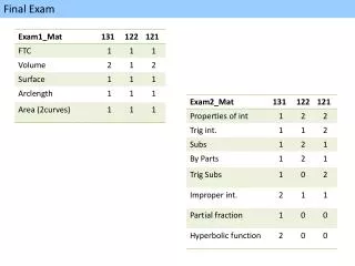
Pulmonary Renal Syndromes Wegener’s Micro AngGoodpasture’s Lupus RPGN Hemoptysis Sinusitis Serology c-ANCA p-ANCA anti-GBM Ab ANA Crescents Necrotizing GN Immune deposits Linear IgG Granular Steroids Cyclophosphamide Plasmapheresis Renal prognosis Good Good Poor Good Lung prognosis Good Good Good Good Pathology Treatment
WEGENER’S GRANULOMATOSIS:Signs and Symptoms • SINUSITIS • Lungs: • Hemoptysis • “Coin lesions” • Crackles/Rales • Kidneys: • RPGN: Crescents • Necrotizing GN • Skin: • Purpura • c-ANCA + serology
WEGNER’S GRANULOMATOSIS All of a sudden, he started coughing up blood (HEMOPTYSIS) And, he didn’t know why, but he was super tired all day (FATIGUE) Corey Anca (C-ANCA) has a really bad sinus infection (SINUSITIS) THEN, a few days later, he got this CRAZY rash (PURPURA) That looked liked some sort of flesh-eating bacteria!
WEGENER’S GRANULOMATOSIS:Pathology • TRIAD of NECROTIZING GRANULOMAS in: • Lungs – acute, URT and LRT • Kidneys – crescentic, focal • Vessels – small–med vessels • MICROSCOPIC FEATURES: • “Geographic” necrosis – irregular • Granulomata – lymphocyes, giant cells, PMNs • Necrotizing vasculitis
WEGENER’S GRANULOMATOSIS:Diagnosis Serology: C-ANCA + UA: 2+ blood, 2+ protein Dysmorphic RBCs = acute nephritis RBC cast = acute nephritis Skin: Purpura = small vessel vasculitis CXR: Dense lesion = granuloma / hemorrhage Lungs: Multiple pale lesions with cavitation Kidney: **Best for tissue diagnosis** Necrosis, Cresent formation NO ELECTRON DENSE DEPOSITS ON ELECTRON MICROSCOPY Artery: Atypical endothelial cells Multinucleated giant cells
WEGENER’S GRANULOMATOSIS:Treatment • Untreated – 90% die within 2 years • Treatment • High dose steroids • Taper over 3-6 months • Cyclophosphamide (2 mg/kg/d) • Continue for 6-12 months after remission • Substitute less toxic drug to complete 12-18 months • Supportive care • Intubation if indicated • Dialysis if indicated
Pulmonary vasculature ** Pulmonary Vascular Disease
ANATOMYPulmonary and Bronchial Arteries Systemic Circulation • Bronchial Arteries Pulmonary Circulation • Originating from the Right Ventricle
Bronchial Circulation • Right Lung 1 artery • Left Lung 2 bronchial arteries • Bronchial arteries “sandwich” the smooth muscle layer in the bronchi • Bronchial arteries also supply the visceral pleura • Bronchial arteries are associated with vascular remodeling of Asthma and COPD
Pulmonary Circulation • Originates from right ventricle • Has thinner walls than systemic arteries • Follows the bronchial tree • Has very sensitive vasoconstrictive properties (hypoxia) • Pulmonary capillaries have a surface area of 125 m2, and make up 85% of alveolar surface area
Cardiac Catheterization What is a Swan-Ganz Catheter? The pulmonary artery catheter allows direct, simultaneous measurement of pressures in the right atrium, right ventricle, pulmonary artery, and the filling pressure ("wedge" pressure) of the left atrium.
Cardiac Catheterization Why do we use cardiac catheters? • To exclude congenital heart disease • To measure wedge pressure or LVEDP • To establish severity and prognosis • To test vasodilator therapy Catheterization is required for every patient with suspected pulmonary hypertension
NORMAL VALUESPulmonary Artery Circulation • Right Atrial Pressure: 2-8 mmHg • Right Ventricle Pressure: 20-30/2-8 mmHg • Pulmonary artery pressure: 25-30/8-15, mPAP = 8-17 mmHg • Pulmonary capillary wedge pressure: 8-15 mmHg • Cardiac Output: 4-6 Liters/min • PVR: change in pressure /flow mPAP-LAp/CO
Interpreting values…Pulmonary Hypertension PVR = mPAP-LAp CO MPAP - mean pulmonary arterial P PCWP/LAp – mean pulmonary artery wedge P Pre-capillary PH PCWP < 15 mmHg PVR > 3 Wu • Pulmonary embolism (PE) • Pulmonary arterial hypertension (PAH) • Respiratory diseases Post-capillary PH PCWP > 15 mmHg PVR normal • Pulmonary vein compression • Atrial myxoma • Mitral valve disease • Myocardial disease • Systemic HTN • Aortic valve disease
PULMONARY EMBOLISM RISK FACTORS (VIRCHOW’S TRIAD) DVT PULMONARY EMBOLISM
PULMONARY EMBOLISM:Clinical Signs and Symptoms • Dyspnea • Pleurisy • Hemoptysis • Syncope • OFTEN SYMPTOMS PRESENT WITH AN EBB AND FLOW, NOT AS A SINGULAR EVENT!! Dangerous because Sx are NON-SPECIFIC! Need to look out for RISK FACTORS and then RULE OUT
PULMONARY EMBOLISM:Effect on Cardiac Physiology Specifically on the right heart • Increased PVR… • Increased RV afterload… • RV failure • Pulmonary embolism: • Reduces the cross-sectional area of the pulmonary vascular bed • Resulting in an increment in pulmonary vascular resistance • Which, in turn, increases the right ventricular afterload. • If the afterload is increased severely, right ventricular failure may ensue.
PULMONARY EMBOLISM:Diagnosis LOW/MODERATE CLINICAL PROBABLITY HIGH CLINICAL PROBABLITY Dyspnea Pleurisy Hemoptysis Syncope EBB & FLOW!! until massive CLINICAL PROBABILITY + D-DIMER CT NON-SPECIFIC SYMPTOMS RISK FACTORS FOR THROMBOSIS • Hypoxia • Unilateral leg swelling • Prior DVT or PE • Recent surgery or trauma (HIP FRACTURES) • Age > 50 • Hormone use • Tachycardia
PULMONARY EMBOLISM:Diagnosis • CXR: normal usually – r/o CHF or rib fracture PE Dx: Hamptons Hump, Westermarks • ECG: ST, right heart strain (sinus tachycardia, R axis deviation, RBBB) • ABG: CO2 up early then decrease • D-Dimer*: Excellent NPV in low suspicion NEG D-DIMER = NOT PE! • Doppler US Extremities: DVT = anticoagulation therapy warrented, used in pregnant women, negative scan does NOT r/o PE • CT PulmAngio: misses small vessels, recommended first line dx imaging • VQ Scan: not useful if CXR is abnormal, used in pregnancy or allergy to contrast • Pulmonary Angiography: *GOLD STANDARD, BUT rarely, if ever used anymore ‘cuz of CT ***PRETEST PROBABILITY***
PULMONARY EMBOLISM:Treatment • Anticoagulation • Heparin first followed by Coumadin • Thrombolysis • tPA, Streptokinase, Urokinase, Alteplase • Vena Caval Filter • Now removable- excellent for trauma wher surgery still planned *** PREVENTS PE, NOT A TX ***
PULMONARY HYPERTENSION PAH: RIGHT HEART
PULMONARY HYPERTENSION:WHO Classifications 1. Primary arterial hypertension • Pulmonary venous hypertension L-CHF • Pulmonary hypertension associated with disorders of the respiratory system and/or hypoxemia OBESITY, SLEEP APNEA, COPD, INTERSTITIAL LUNG DZ • Pulmonary hypertension due to chronic thrombotic and/or embolic disease THROMBOPHILIC DISORDERS • Pulmonary hypertension due to disorders directly affecting the pulmonary vasculature TRAUMA, SARCOIDOSIS, SCHISTOSOMIASIS ATERIAL VENOUS HYPOXIC THROMBO-EMBOLIC MISCELLANEOUS
PULMONARY HYPERTENSION:Difference between PAH and non-PAH IMPORTANT FOR DETERMINING TREATMENT!!
PULMONARY ARTERIAL HYPERTENSION:Predisposing Factors • Family history • Sleep apnea • Heart murmur • Arthralgias, arthritis, rash • Liver disease • Appetite suppressant exposure • Deep venous thrombosis or pulmonary embolism • HIV risk factors • Underlying lung disease • Congenital heart disorders
PULMONARY ARTERIAL HYPERTENSION:Predisposing Features Pulmonary Arterial Hypertension 1. Primary pulmonary hypertension (a) sporadic – YOUNG WOMEN, 20-30’S (b) familial – BMPR RECEPTOR 2. Related to: (a) collagen vascular disease - SCLERODERMA (b) congenital systemic to pulmonary shunts – ASD, VSD, PDA (c) portal hypertension (d) HIV infection – 1/200 PTS (e) anorexigens - METHAMPHETAMINES (f) persistent pulmonary hypertension of the newborn – PREMATURE BABIES
PULMONARY HYPERTENSION:Clinical Signs and Symptoms • Characterized by progressive elevation of pulmonary artery pressure and vascular resistance • Patients are limited by: • Shortness of breath • Dyspnea on exertion • Pre-syncope and syncope • Chest pain • Edema and ascites • Often leads to right ventricular failure and death • Jugular venous distention • Right ventricular heave • Right-sided fourth heart sound • Loud pulmonic valve closure (P2) • Peripheral edema, ascites
PULMONARY HYPERTENSION:Diagnosis NOTE: CATHETERIZATION is required for every patient with suspected pulmonary hypertension D DOPPLER ECHOCARDIOGRAM To see RVSP, RAE/RVE, pericardial effusion, and estimate systolic PAP RIGHT VENTRICULAR dysfunction D-SIGN IF PAH IS LIKELY…
PULMONARY ATERIAL HYPERTENSION:Treatment • Anticoagulation • Diuretic • Digoxin – MOST POTENT VASODILATOR • Oxygen • Vasodilators • CCB • Endothelin Blocker • Sildenafil • Prostacyclins • Nitric oxide
PULMONARY ARTERIAL HYPERTENSION:Histopathological Findings Increased pressures cause thickening of the intima and fibrosis over time… MEDIAL HYPERTROPHY INTIMAL HYPERPLASIA (OCCLUSION)
RESPIRATORY FAILURE A clinical syndrome in which the respiratory system is unable to adequately meet the body’s demands for CO2 elimination or oxygen delivery
RESPIRATORY FAILURE:Signs and Symptoms CLINICAL DIAGNOSIS! • Shortness of breath • Rapid breathing (tachypnea) • Air hunger • Nasal flaring • Use of accessory muscles • Paradoxical respirations • Severe: • Bluish color on your skin, lips, and fingernails • Confusion • Sleepiness
RESPIRATORY FAILURE:Diagnosis • CLINICAL diagnosis! • Role of ABG:Not necessary!! • Can be helpful for distinguishing Type 1 vs. Type 2
RESPIRATORY FAILURE:Treatment • The thoughtful approach…. • You have determined that your patient has respiratory failure, you have distinguished between type I and II RF, and you have determined why this patient has respiratory failure. • Now use your understanding of physiology and disease to target therapies to correct the abnormal physiology and disease. • The “I don’t have time to be thoughtful” approach…. • Stabilize your patient – airway, hemodynamics, oxygenation, and ventilation. • Let the dust settle, and return to approach #1.
RESPIRATORY FAILURE:Type I Treatment IMPROVE OXYGENATION Increase Oxygenation: Increase inspiratory pressure Increase FiO2
RESPIRATORY FAILURE:Type II Treatment IMPROVE VENTILATION Increase Ventilation: Decrease I time (increase E) Allow for permissive hypercapnea or tracheal gas insufflation
RESPIRATORY FAILURE:Mechanical Ventilation • What problem is the ventilator reversing?... • From paper… • Objectives of Mechanical Ventilation • Improve pulmonary gas exchange • Reverse hypoxemia • Relieve acute respiratory acidosis • Relieve respiratory distress • Decrease oxygen cost of breathing • Reverse respiratory-muscle fatigue • Alter pressure-volume relations • Prevent and reverse atelectasis • Improve compliance • Prevent further injury • Permit lung and airway healing • Avoid complications
ACUTE RESPIRATORY DISTRESS • Non-cardiogenic, pulmonary edema resulting from severe acute alveolar injury • Four main criteria for ARDS: • Hypoxia • Chest X-Ray: Bilateral diffuse infiltrates of the lungs • No cardiovascular lesion • PF ratio is less than 200
ARDS:Clinical Signs and Symptoms • Shortness of breath • Tachypnea • Confusion • Occurs within 24-48 hours of an injury or an acute illness • SIGNS: • Bilateral infiltrates on CXR sparing costophrenic angles • PAWP < 18 mmHg • PaO2: FiO2 < 200 mmHg ACUTE SYNDROME THAT AFFECTS THE LUNGS WIDELY AND RESULTS IN SEVERE OXYGENATION DEFECT THAT IS NOT DUE TO HEART FAILURE!
ARDS:Treatment and Therapies • Mechanical ventilation • APRV • PEEP • Prone position • Relieves atelectasis • Fluid management • Diuresis • Fluid restriction • Corticosteroids • Modest dose Methylprednisolone • NO • Surfactant therapy
ARDS: Mechanical Ventilation End inspiratory pause Decelerating wave form Flow Pressure Plateau pressure Volume Time