Download
1 / 56
580 likes | 995 Views
SYSTEMIC LUPUS ERYTHEMATOSUS (SLE). SYSTEM IC LUPUS ERYTHEMATOSUS (SLE) Gergely Péter dr.

E N D
SYSTEMIC LUPUS ERYTHEMATOSUS (SLE) SYSTEMIC LUPUS ERYTHEMATOSUS (SLE) Gergely Péter dr Definition:The clinical picture of SLE is variable. The most common form of the disease: young female with non-erosive arthritis, (low grade) fever, elevated ESR, leucopenia (with or without skin and kidney involvement). Epidemiology:Incidence: 0.7/100,000, prevalence 23-50/100,000. In Hungary the expected number of SLE cases may be between 2500 and 5000. Female dominance (male: female ratio: 1:9).Familial aggregation is known.
Pathomechanism of SLE Endogeneous factors: genetic hormonal Exogeneic factors: infections, UV radiation drugs (hydralazine, INH, quinidine, procainamide) B cell hyperreactivity enhanced autoantibody production, in particular antinuclear antibody (ANA) production immune complex formation and deposition tussue inflammation (skin, joints, kidney) organ lesion/regeneration
Lupus band test = IC deposition in the skin Lupus band test = IC deposition in the skin
ANA ANA
ANA ANA
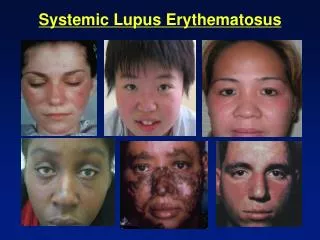
Clinical features of SLE Clinical picture: The well known butterfly (malar) rash is relatively uncommon, but the diagnosis is in most cases easy, if we base it on the criteria of American Rheumatism Association (ARA). General symptoms: fever, lymphadenomegaly, weight loss are always present, their severity depends on the intensity of the disease. At first sight, such patient seem to be suffering of an infectious disease.
Joints Joints: are frequently involved, moderate swelling and morning stiffness are the dominant features. Unlike in RA, no erosions and deformity is seen. Subluxation and Jaccoud type arthropathy are rare forms of SLE.
Jaccoud arthropathy in SLE Jaccoud arthropathy in SLE
Skin involvement is frequent Skin involvement is frequent. Beside butterfly and discoid rash, chronic (IC-mediated) urticaria, livedo reticularis, skin vasculitis, biphasic Raynaud’s phenomenon, hair thickening and alopecia areata may be present.
Malar (butterfly)rash in SLE Malar (butterfly) rash in SLE
Malar rash in SLE Malar rash in SLE
Malar rash in SLE Malar rash in SLE
Discoid lesion Discoid lesion
Chronic DLE on the face Chronic DLE on the face
Discoid on the scalp Discoid on the scalp
DLE on the scalp DLE on the scalp
SCLE SCLE
SCLE SCLE
Raynaud phenomenon Raynaud phenomenon
Perinugual vasculitis Perinugual vasculitis
Perinugual vasculitis Periungual vasculitis
Vasculitis on the feet Vasculitis on the feet
Livedo reticularis Livedo reticularis
Lupus pernio Lupus pernio
Renal involvement is observed in 60% of patients Renal involvement is observed in 60% of patients. SLE may manifest as a monosystemic glomerulonephritis, but usually other organs are also involved. Glomerulonephritis is an IC-mediated inflammation with various histological and clinical signs: from mild proteinuria to rapidly progressive glomerulonephritis. Nephrosis syndrome is very common. According to histology, nephritis can be classified. WHO classification is shown below (in parentheses: clinical manifestations) I. normal (no alterations) II. mesangial (mild proteinuria) III. focal segmental (marked proteinuria, positive sediment) IV. diffuse (acute nephritis or nephrosis) V. membranous (nephrosis) VI. sclerosis/end stage.
Generalized edema in nephrotic syndrome Generalized edema in nephrotic syndrome
Lupus nephritis (WHO I) Lupus nephritis (WHO I)
Lupus nephritis (WHO II) Lupus nephritis (WHO II)
Lupus nephritis (WHO III) Lupus nephritis (WHO III)
Lupus nephritis (WHO IV) Lupus nephritis (WHO IV)
IC deposition in the glomeruli - SLE IC deposition in the glomeruli - SLE
Hematological disorders are very common Hematological disorders are very common. A mild anemia (anemia of chronic disease) is always almost present in active disease, frank hemolysis is rare. Leucopenia is common, but even patients with leukocyte count <2.0 have no problems at all. Lymphopenia can be profound (0.5), granulocytopenia rarely results in infections. Thrombocytopenia is more common in patients with APS. Central nervous system involvement is common, in particular in patients with APS. One of the most reliable method to detect CNS involvement is MRI (however, some foci, seen in MRI may be asymptomatic, therefore the correlation between MRI changes and clinical picture is weak). Psychosis due to corticosteroid treatment is much less frequent that caused by the disease itself. In cuch cases, when in doubt, a bolus corticosteroid is more likely of benefit then withdawal. There are also minor signs: headache, cognitive and emotional disturbances, e.d. depression, etc.
ACR criteria of CNS involvement ACR criteria of CNS involvement: acuteinflammatory demyelinizing polyradiculopathy (Guillain-Barré syndrome) aseptic meningitis autonomousnervous system alterations (e.g. orthostatic hypotension) cerebrovascular disease (e.g. stroke) demyelinization syndrome lupus headache mononeuropathy myasthenia gravis cranial neuropathy plexopathy convulsions acute confusionstate anxiety cognitive dysfunction mood disorders
Multiple infarcerationsin SLE –APS (MRI) Multiple infarcerations in SLE –APS (MRI)
Antiphospholipid syndrome (Hughes’s syndrome) Antiphospholipid syndrome (Hughes’s syndrome) A relatively recently decribed syndrome characterized by recurrent venous (or arterial) thromboses, thrombocytopenia, stroke, and in women, recurrent fetal loss. Less frequently other thromboembolic complications may also occur. The syndrome is caused by autoantibodies directed against phospholipid – glycoprotein complexes interfering with normal clotting mechanism.
Diagnostic criteria of SLE (ARA, 1982, modified in 1997) • Diagnostic criteria of SLE (ARA, 1982, modified in 1997) • Malar rash (or: vespertilio, butterfly rash) • Discoid rash • Photosensitivity • Oral ulcers: oral or nasopharyngeal ulceration • Arthritis: nonerosive athritis • Serositis: (at least one of the following) • a) pleuritis • b) pericarditis • 7. Renal disorder (at least one of the following): • a) persistent proteinuria (>0.5g/day or 3+) • b) cellular casts (erythrocyte, hemoglobin, granular, tubular or mixed) • 8. Neurological disorder (at least one of the following): • a) a) seizures • b)b) psychosis • 9. Hematologic disorder (at least one of the following): • a) a) hemolytic anemia (with reticulocytosis), or • b) b) leukopenia (<4.0 on 2 or more occasions), or • c) c) lymphopenia (<1.5 on 2 or more occasions), or • d) d) thrombocytopenia (<100 in the absence of offending drug) • 10. Immunologic disorder (at least one of the following): • a) a) abnormal titer anti-dsDNA antibody, or • b) b) antibody to Sm nuclear antigen, or • c) c) abnormal titer of anticardiolipin antibody • ANA positivity • If 4 criteria are present serially or simultaneously, the diagnosis=SLE
Autoantibodies in SLE Autoantibodies in SLE Antigen NatureFrequency (%) Native DNA (double-stranded DNA) 40 Denatured DNA (single-stranded DNA) 70 Histon H1, H2A, H2B, H3, H4) 70* Sm RNA-protein complexes 30 nuclear-RNP (U1-nRNP) 32 SS-A (Ro) RNA-protein complexes 35 SS-B (La) RNA-protein complexes 15 Ku protein 10 ribosomal-RNP phosphoproteins 10 Ki/S1 protein 6 * >90% in drug-induced SLE
LE cell phenomenon LE cell phenomenon
Homogenous ANA positivity on HEp-2 cells Homogenous ANA positivity on HEp-2 cells
Peripheral ANA positivity Peripheral ANA positivity
Speckled ANA positivity Speckled ANA positivity
Nucleolar ANA positivity Nucleolar ANA positivity
Anti-DNA positivity (Crithidia luciliae test) Anti-DNA positivity (Crithidia luciliae test)
Differential diagnosis Differential diagnosis. In all cases SLE should be clearly differentiated from: 1. other systemic autoimmune diseases 2. infections 3. malignancies, especially lymphomas.
The activity of the disease (monitoring)can be assessed by the followings The activity of the disease (monitoring) can be assessed by the followings: 1. Clinical signs and symptoms are of utmost importance. 2. Blood count: leukopenia cannot be used, but thrombocyto-penia andhemolysis are of importance. 3. ESR may reflect disease activity, high valueusually indicates acivity, but it remains high in many cases in remission. 4. Renal functions (serum creatinine) and proteinuria should be checked regularly. High creatinine and an increase in daily proteinuria usually indicate an activation. 5. Anti-DNA level runs parallel with activity, but it may remain elevated in remission. 6. Complement activity and C3, C4 levels also indicate activity, they decrease during activity, and tend to normalize in remission. 7. CRP usually remains normal, an increase may indicate bacterial infection. 8. There are indices to measure overall disease activity, e.g. SLEDAI (SLE disease activity index).
SLEDAI SLEDAI ScoreSymptom 8 acute convulsion 8 psychosis 8 "organic brain" syndrome 8 visual disturbances (retinal vasculitis) 8 cranial nervesign (sensory, motor) 8 lupus cephalalgia 8 cerebrovascular laesion (excluded: atherosclerosis) 8 vasculitis (ulcus, gangrena, skin vasculitis) 4 myositis 4 arthritis (> 2 joints) 4 granular or erythrocyte casts 4 hematuria (>5 red cells/field) 4 new proteinuria (>0.5 g/day), or >+0.5g/dayincrease 4 pyuria (>5 leukocytes/field) (excluded: infection) 2 new exanthema 2 alopecia, newor recurrent 2 mucous membrane ulcer 2 pleuritis 2 pericarditis 2 lowcomplement (CH50, C3, etc.) 2 high anti-DNA 1 fever (>38oC) (excluded: infection) 1 thrombocytopenia (<100,0) 1 leucopenia (<3,0) (wxcluded: drug-induced) TOTAL SLEDAI SCORE
Therapy of SLE • Therapy of SLE • 1) Inactive • Check-up: 3 months: blood count, urine, creatinine, • blood pressure, once a year: immunology (anti-DNAcomplement, anti-cardiolipin etc. • 2) moderately active (complaints, laboratory abnormalities, no general signs) • Organ involvement: • a) skinand/orjointsand/ormoderate serositis • NSAID and/or(hydroxy)chloroquine • RESPONSE continue chloroquine for at least 6 mo(fundus control after 2 months, then every 6 months) • NSAID as required • NO RESPONSE 30 mg prednisolone/day (24 mgmethyl- • prednisolone) for 1 week, then slowly (during 2 weeks) • taper to 5-10 mg maintenance dose; • discontinue if possible. • OR: 7.5 mg methotrexate (MTX) per week (up to 15 • mg/week)
hematology • b) hematology: • leucopenia: no treatment • anemia: only hemolysis should be treated if HTC<0.3 • hemolysis: 30 mg or more prednisolone/day, maintenance • dose, and/or MTX added • thrombocytopenia: if >50: no treatment if <20 prednisolone 30 mg or more/day, then maintenance dose • if instable: danazole, vincristinec) kidney: • normalcreatinine, moderate (0.2-1 g/day) proteinuria, minimal sediment; if stable only observation, biopsy = treatment options according to WHO grade. • d) ACL positivity without clinical APS:only observation