Download
1 / 21
220 likes | 491 Views
The III nerve palsy: when to panic. UBC Clinical neuroophthalmology day - 18 November 2005 Jason Barton. ?. III nerve palsies. Nuclear III palsy: bilateral superior rectus weakness, bilateral ptosis, bilateral pupil paresis .

E N D
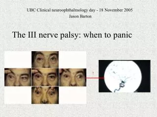
The III nerve palsy: when to panic UBC Clinical neuroophthalmology day - 18 November 2005 Jason Barton ?
Nuclear III palsy:bilateral superior rectus weakness, bilateral ptosis, bilateral pupil paresis
39 year old man with HIV/AIDS, presenting with headache and bilateral ptosis. Exam showed limited downgaze od more than os, and bilateral ptosis. Autopsy showed HIV encephalitis surrounding the aqueduct.
Fascicular III palsy: may mimic divisional palsies, monocular elevation paresis, or isolated paresis of the pupil or inferior oblique ipsilateral limb ataxia (brachium conjunctivum) contralateral resting tremor/chorea/ballismus (red nucleus) contralateral paresis of the limbs and/or face (cerebral peduncle) vertical supranuclear palsy (riMLF, inC) drowsiness Causes of midbrain III syndromes: vascular 'top of the basilar' syndromes. intrinsic primary or secondary tumors infections (AIDS, TB) demyelination brainstem hemorrhages (including uncal herniation)
Bilateral fascicular III palsies 68 year old man with hypertension, awoke with bilateral ptosis. Exam: 0% elevation and 0% depression ou, 50% adduction od and 0% adduction os. Severe ptosis: 6mm lid fissure od and 4mm os, with frontalis contraction. Pupils 4mm od and 2.5mm os, with light reaction. Left leg paresis. MRI with DWI
Peripheral III palsy: subarachnoid portion contralateral hemiparesis. Meningismus depending on etiology. Causes: Aneurysms: basilar or ICA-pcomm artery Ischemia: hypertension/DM. sildenafil/cocaine, GCA, carotid occlusion. Trauma: LOC or skull fractures, uncal herniation Tumors: neurinomas and schwannomas Inflammation: Lyme, sarcoidosis - rare.
Woman anticoagulated for atrial fibrillation with right temporal lobe and capsular stroke, returned 1 year later with new right III palsy. 1.5 years later, showed signs of aberrant regeneration also (elevation accompanied by adduction).
Peripheral III palsy: Cavernous sinus lesions: divisional paresis IV V and VI, but can cause III mononeuropathies. Causes: Vascular: dural arteriovenous malformations, giant ICA aneurysm Tumours: 1° - meningioma, chordoma, craniopharyngioma, 2° - nasopharyngeal ca Vascular/tumour: pituitary apoplexy ! Inflammation: viral or flu-like infections, sphenoid sinusitis
Aneurysms: basilar or ICA-pcommA aneurysms: usually acute painful palsy. “Natural history” - Soni, JNNP 1974 • rupture of ICA-PCA aneurysms preceded by warning sign 70% of time. • average time between initial symptoms and subarachnoid hemorrhage = 30 days. • delay in treating ICA-PCA aneurysm increases mortality from 20% to 67%.
Best way to detect an aneurysm? (Can we avoid 1-2% risks of formal angiography?) • CTA and MRA detect aneurysms of ≥ 4-5mm. Is that good enough? -Yanaka et al, Neurosurg 2003: aneurysms of 4-8mm can present with III palsy -Jacobson & Trobe, AJO 1999: MRA would miss < 1.5% of aneurysms with III palsy • Is the prognosis of small ICA-PCA aneurysms with III palsy better than large ones? For asymptomatic aneurysms, risk of SAH ≈ size and location. NEJM 2004: <10mm = 0.05%/yr, pcomm relative risk = 8X that of other aneurysms. “…unlikely that surgery will reduce the rates of disability and death in patients with unruptured aneurysms smaller than 10mm in diameter and no history of subarachnoid hemorrhage.” • Unknown: does this same conclusion hold for ICA-PCA aneurysms with symptoms from compression of III nerve rather than SAH?
Ischemic palsies: mean age of 61 years microvascular palsy presents as a sudden onset painful III nerve defect with frequent sparing of the pupil. • in many, diabetes is discovered after the III nerve palsy. • time course: 1. Some progression is commonly in the first 1-3 weeks. 2. Pain should not last more than 7-14 days. If it does, worry. 3. Recovery 1 to 3 months later. If not, image for tumors/aneurysms. 4. Spontaneous recovery can happen after weeks/ months with p-comm aneurysms and lymphoma. Recovery with compression or trauma can even occur after several years.
Three rules to remember: The pupil rule: “complete pupil sparing with complete EOM involvement is not going to be from an aneurysm” (after Lee et al, Survey of Ophthalmology 2002)
66 year old man with longstanding DM, pupil-sparing complete III palsy, with pain for 1 week. Recovered after 3 months.
Three rules to remember: The pupil rule: complete pupil sparing with complete EOM involvement is not going to be from an aneurysm. Aberrant regeneration - implies compression until proven otherwise
III nerve palsy with aberrant regeneration cavernous sinus meningioma
Three rules to remember: The pupil rule: complete pupil sparing with complete EOM involvement is not going to be from an aneurysm. Aberrant regeneration - implies compression until proven otherwise Divisional palsies - think cavernous sinus mass
Inferior divisional III palsy
III nerve palsy summary: 1. Can you locate the site of damage? (i) Does it localize a nuclear site? (ii) Are there neighbourhood neurologic signs? 2. Do the three rules suggest an aneurysm or mass? (i) pupil rule (ii) aberrant regeneration rule (iii) divisional palsy rule If it is suspicious, MRI/CT sella with contrast, CTAngiogram If negative but high index of suspicion, formal angiogram, and don’t forget CSF in appropriate cases