Extra Slides Orientation Images
140 likes | 227 Views
Explore the detailed anatomy of the lung, including trachea, bronchus, pulmonary artery, alveoli, and different cell types. Learn about respiratory epithelium, elastic fibers, and pulmonary veins.

Extra Slides Orientation Images
E N D
Presentation Transcript
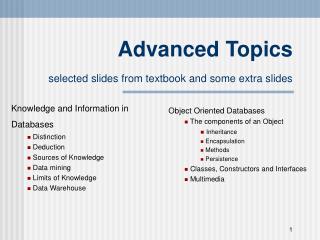
#40; Trachea Basement membrane Respiratory epithelium Elastic fibers
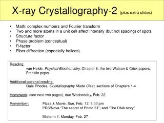
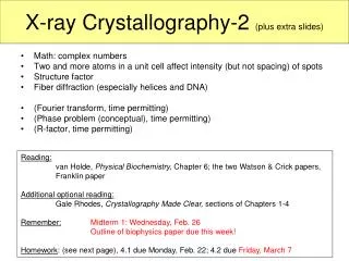
#130-1 Lung 1? Bronchus 2? Pulmonary artery
#130-1 Lung Pulmonary artery Close to bronchial trees relatively thin wall (media) elastic laminae relatively wide lumen
#130-1 Lung 4? 3? Bronchial vein Bronchial artery Only in the wall of large bronchi. Ordinary arteries with relatively thick media. Bronchial cartilage
#130-1 Lung Bronchiole Ciliated cells non-ciliated cells Ciliated and non-ciliated (Clara) cells No goblet cells Prominent sm. M. Smooth muscle
#132-2 (Terminal) Bronchiole & Pulmonary artery Smooth Muscle P. A. Bronchiole
#132Lung 7? P.A. 6? Respiratory bronchiole 5? Terminal bronchiole Low cells alveolar pocketing Knobs of sm. muscle (arrows)
#129 Lung Alveolar duct 8? alveoli 9?
#130-1 Lung 10? 12? Type I Pneumocyte Macrophage 11? Type II Pneumocyte (surfactant)
#130-1 Lung Away from bronchial trees thin wall Pulmonary vein 13?
#130-2 Lung Pulmonary vein
#129 Lung ? ? ?
Slide 42: Lung, chronic congestion – hemosiderin-laden macrophages lung alveolar air space hemosiderin-laden macrophages