Download
1 / 17
170 likes | 368 Views
AS Biology Core Principles. The Electron Microscope. Aims. Resolving power The resolving power of light & electron microscopes The difference between the light & electron microscope Transmission & scanning electron microscopy. Introduction. Microscopes magnify & resolve images

E N D
AS Biology Core Principles The Electron Microscope
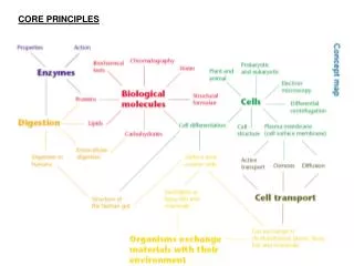
Aims • Resolving power • The resolving power of light & electron microscopes • The difference between the light & electron microscope • Transmission & scanning electron microscopy
Introduction • Microscopes magnify & resolve images • Microscopy began in 1665 when Robert Hooke coined the word ‘cells’ to describe the structure of cork • You need to know about 2 types of microscope - light & electron • You need to know how they work and the differences between them • ‘Its not how much they magnify that is key - but how well they resolve…’
Resolving Power • The limit of resolution of a microscope is the smallest distance between 2 points that can be seen using a microscope • This is a measure of the clarity of the image • A microscope with a high resolving power will allow 2 small objects which are close together to be seen as 2 distinct objects
Resolving Power • Resolving power is inversely proportional to the wavelength of the radiation it uses
The Light Microscope • Series of lenses through which ordinary white light can be focused • Optical microscopes can not resolve 2 points closer together than about half (0.45) the wavelength of the light used (450-600nm) • How close is this?
The Light Microscope • The total magnification is the eyepiece magnification multiplied by the objective magnification • The maximum magnification of a light microscope is x1500 • What can it be used for? • What can it not be used for?
The Electron Microscope • Electrons (negatively charged, very small particles) can behave as waves • The wavelength of electrons is about 0.005nm • What will this mean for the limit of resolution? • Electrons are ‘fired’ from an electron gun at the specimen and onto a fluorescent screen or photographic plate • Where is this technique commonly used? • There are 2 types of electron microscopy - transmission and scanning • Both focus an electron beam onto the specimen using electromagnets
Transmission Electron Microscope (TEM) • In transmission EM the electrons pass through the specimen • Specimen needs to be extremely thin - 10nm to 100nm • TEM can magnify objects up to 500 000 times • TEM has made it possible to see the details of and discover new organelles - see page 9 in Collins
Transmission Electron Microscope (TEM) • Cells or tissues are killed and chemically ‘fixed’ in a complicated and harsh treatment (in full detail in table 3.1 pg 52 Rowland) • How does this differ to light microscopy? • This treatment can result in alterations to the cell - known as artefacts • What will this mean for the images produced?
Transmission Electron Microscope (TEM) Transmission electron micrograph of epithelial cells from a rat small intestine. Scale bar = 5 mm.
Scanning Electron Microscope (SEM) • In Scanning EM microscopes the electrons bounce off the surface of the specimen • Produce images with a three-dimensional appearance • Allow detailed study of surfaces
Scanning Electron Microscope (SEM) Now watch the following clip explaining SEM
Links • www.learn.co.uk/ • www.microscopy-uk.org.uk/intro/index.html • www.mwrn.com/feature/education.asp • http://www.feic.com/support/tem/transmis.htm • http://anka.livstek.lth.se:2080/microscopy/foodmicr.htm
Light & Electron Microscopes Copy & complete the following table
This powerpoint was kindly donated to www.worldofteaching.com http://www.worldofteaching.com is home to over a thousand powerpoints submitted by teachers. This is a completely free site and requires no registration. Please visit and I hope it will help in your teaching.