Download
1 / 74
740 likes | 1k Views
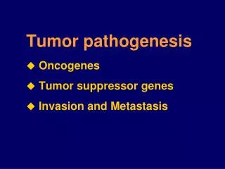
Tumor pathogenesis. 陈玮 副教授 Email : chenwei566@zju.edu.cn 个人主页: http://mypage.zju.edu.cn/566 8888. Tumor pathogenesis Oncogenes Tumor suppressor genes Invasion and Metastasis. Introduction.

E N D
Tumor pathogenesis 陈玮 副教授 Email:chenwei566@zju.edu.cn 个人主页: http://mypage.zju.edu.cn/566 8888
Tumor pathogenesis • Oncogenes • Tumor suppressor genes • Invasion and Metastasis
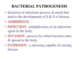
Introduction • Carcinogensis is multistep process involving the multiple genetic changes including the activation of cooperating oncogenes and the inactivation of tumor suppressors in somatic cells 3
Molecular alterations during human colon tumor progression (~ 40-50 %) (~ 60 %) * (~ 90 %) * (~ 50-70 %) The precise contribution of hypomethylation to tumor progression remains unclear; some evidence suggests that it creates chromosomal instability.
Usually, a single oncogene is not enough to turn a normal cell into a cancer cell, and many mutations in a number of different genes may be required to make a cell cancerous.
Figure 2. Intracellular Signaling Networks Regulate the Operations of the Cancer Cell. An elaborate integrated circuit operates within normal cells and is reprogrammed to regulate hallmark capabilities within cancer cells. Separate subcircuits, depicted here in differently colored fields, are specialized to orchestrate the various capabilities. At one level, this depiction is simplistic, as there is considerable crosstalk between such subcircuits. In addition, because each cancer cell is exposed to a complex mixture of signals from its microenvironment, each of these subcircuits is connected with signals originating from other cells in the tumor microenvironment, as outlined in Figure 5. (Hanahan D, Weinberg RA. Hallmarks of Cancer: The Next Generation. Cell 2011, 144:646)
Oncogene Concept: An oncogene is a gene that when mutated or expressed at abnormally-high levels contributes to converting a normal cell into a cancer cell. • Cellular oncogene (c-onc): --- proto-oncogene (proto-onc):in normal physiologic version --- Oncogene:altered in cancer • Viral oncogene (v-onc)
Fuctions of proto-oncogenes • Proto-oncogenes have been identified at all levels of the various signal transduction cascades that control cell growth, proliferation and differentiation: • extracellular proteins function as growth factors, • membrane proteins as cell surface receptors • cellular proteins that relay signals • proteins innucleus, which activate the transcription and promote the cell cycle • This signaling process involves a series of steps that: • begin from the extracellular environment to cell membrane; • involve a host of intermediaries in the cytoplasm; • end in the nucleus with the activation of transcription factors that help to move the cell through its growth cycle.
Classification of proto-oncogenes • Growth factors, e.g. V-sis (PDGF), int-2 (FGF) • Receptor Tyrosine Kinases,e.g.Her-2/ neu/ erbb2 (EGFR) • Membrane Associated Non-Receptor Tyrosine Kinases, e.g.src, Lck • Membrane Associated G-Proteins,e.g. Ras • Serine/Threonine Kinases e.g. Raf • Nuclear DNA-Binding/Transcription Factors, e.g. myc, fos • Others • Apoptosis regulators, e.g. Bcl-2, • Regulators of cell cycle, e.g.Cyclin D1, CDK4
Mechanisms of Oncogene Activation • Gene amplification, e.g.myc, CCND1 • Point mutation, e.g.ras, • Chromosomal rearrangement or translocation • the transcriptional activation of proto-onc. • the creation of fusion genes, e.g.abl-bcr • Viral insertion activation, e.g.c-Myc
Amplification Translocation
CHROMOSOMAL REARRANGEMENTS OR TRANSLOCATIONS Neoplasm Translocation Proto-oncogene Burkitt lymphoma t(8;14) 80% of cases c-myc1 t(8;22) 15% of cases t(2;8) 5% of cases Chronic myelogenous t(9;22) 90-95% of cases bcr-abl2 leukemia Acute lymphocytic t(9;22) 10-15% of cases bcr-abl2 Leukemia 1c-myc is translocated to the IgG locus, which results in its activated expression 2bcr-abl fusion protein is produced, which results in a constitutively active abl kinase
GENE AMPLIFICATION Oncogene Amplification Source of tumor c-myc ~20-fold leukemia and lung carcinoma N-myc 5-1,000-fold neuroblastoma retinoblastoma L-myc 10-20-fold small-cell lung cancer c-abl ~5-fold chronic myeloid leukemia c-myb 5-10-fold acute myeloid leukemia colon carcinoma c-erbB ~30-fold epidermoid carcinoma K-ras 4-20-fold colon carcinoma 30-60-fold adrenocortical carcinoma
Ras • Locates on chromosome 11, codes for a protein with GTPase activity • relays signals by acting as a switch:When receptors on the cell surface are stimulated, Ras is switched on and transduces signals that tell the cell to grow. If the cell-surface receptor is not stimulated, Ras is not activated and so the pathway that results in cell growth is not initiated. • mutated in about 30% of human cancers so that it is permanently switched on, telling the cell to grow regardless of whether receptors on the cell surface are activated or not.
Ras relays signals from the cell surface receptors to the nucleus Ras relays signals by acting as a switch
Her2/neu/erbB-2 • This gene was discovered by three different groups. That is why it has three different names. • It is a member of EGFR superfamily, also be a receptor tyrosine kinases • Dr. Slamon (UCLA) described the role of Her2/neu in breast cancer and ovarian cancer. • Overexpression, amplification, rare translocations • No ligand is known
Prospect • A breakthrough for our understanding of the molecular and genetic basis of cancer • Provided important knowledge concerning the regulation of normal cell proliferation, differentiation, and programed cell death. • The identification of oncogene abnormalities has provided tools for the molecular diagnosis and monitoring of cancer. • Oncogenes represent potential targets for new types of cancer therapies.
Tumor suppressor genes Concept: genes that sustain loss-of-function mutations in the development of cancer
TSGs Transcriptional factor: p53, WT1, Direct transcription regulator: Rb, APC Inhibitor of cell cylcle kinase: p16INK4A, p19ARF, Cell structural components: NF2 Phosphatase: PTEN Potential mediator of mRNA processing: VHL Components involved in DNA repair: MSH2, MLH1, BRCA1, p53
TUMOR SUPPRESSOR GENES Disorders in which gene is affected Gene (locus) Function Familial Sporadic DCC (18q) cell surface unknown colorectal interactions cancer WT1 (11p) transcription Wilm’s tumor lung cancer Rb1 (13q) transcription retinoblastoma small-cell lung carcinoma p53 (17p) transcription Li-Fraumeni breast, colon, syndrome & lung cancer BRCA1(17q) transcriptional breast cancer breast/ovarian tumors BRCA2 (13q) regulator/DNA repair
Mechanism for the inactivation of TSGs • Mutation: point mutation or frameshift mutation, p53 • Deletion: LOH (loss of heterozygosity) or homozygous deletion, Rb • Viral oncoprotein inactivation: p53, Rb • Promoter hypermethylation, histone modification changes: p16
Rb regulates G1/S transition Rb inactivation by viral oncoprotein
RB • Retinoblastoma is an uncommon childhood tumor • Retinoblastoma is either sporadic (60%) or familial ( 40% ) • Two mutations required to produce retinoblastoma • Both normal copies of the gene should be lost to produce retinoblastoma
RB RB RB LOH RB Mutation KNUDSON TWO HIT HYPOTHESIS IN SPORADIC CASES Normal Cells RB RB Inactivation of a tumor suppressor gene requires two somatic mutations. Tumor cells
P53: • Common in human cancer 70% • Tumor suppressor gene • DNA damage – P53 activation by release from MDM2 • P53 results in arresting the cell cycle by increasing P21. • P53 enhance repair of the DNA damage by GADD45 • P53 induces apoptosis by increasing level of Bax
Bax P53 Function as gatekeeper • Inactivation of p53 in cancer • LOH on 17p13 in a number of tumors • Point mutation on exon 5-8 “hot-spot” (Dominant negative mutation) • MDM2 negative regulation • viral-oncogene products inactivation
P53 is called the “ guardian of the genome” • 70% of human cancers have a defect in P53 • It has been reported with almost all types of cancers : e.g. lung, colon, breast • In most cases, mutations are acquired, but can be inhereted, e.g : Li-Fraumeni syndrome
PTEN (Phosphatase and Tensin Homolog) • Pten gene located on Chrom 10 (10q23) • PTEN was discovered in 1997 as the first tumor suppressor phosphatase • PTEN and PI-3 Kinase act as antagonists in lipid signaling
PTENis a lipid 3-phosphatase, which signals down the PI3 kinase/AKT proapoptotic pathway. Backman et al.
Outcomes of the Akt/PKB pathway • Role in proliferation complicated • PTEN does not merely block proliferation because studies showed that normal bacteria cells expressing PTEN can still undergo rapid proliferation (Liliental et al., 2000) • Role in apoptosis more clear • Re-expression of PTEN in several carcinoma cell lines induces apoptosis (Li et al, 1998) • Especially important is Anoikis, a form of apoptosis that occurs when cells lose contact with the e.c.m. -mediates the cell’s “anchorage dependence” (Davies et al., 1998)
How is Pten involved in cancer? • Most frequently mutated gene identified yet in endometrial cancers (33-55% of tumors examined) • Of 647 Malignant Glial Tumors examined, 24% showed mutations in PTEN • Ovarian tumors (of endometriod origin) showed mutations in 26% of tumors • Prostate Carcinoma-mutations in 18% of tumors
The process of metastasis consists of sequential linked steps • Growth at primary site and angiogenesis • Tumor cell invasion • Lymphatic and hematogenous metastasis • Growth at secondary site and angiogenesis
Mechanisms involved in tumor cell invasion 1.Loss of cell-to cell cohesive forces 2. Secretion of ECM-degrading enzymes 3. Active Locomotion 4. Tumor angiogenesis 5. Metastasis-related genes
Degradation of ECM by collagenase enzyme Detachment of tumor cells from each other Attachments of tumor cells to matrix components Migration of tumor cells
5. Metastasis-enhancing Genes:Oncogenes,CD44, Integrinβ1, CEA, MMP2, u-PA, etc
1. Loss of cell-to cell cohesive forces: • Cell adhesion molecules (CAMs): • 细胞粘附分子:介导细胞之间或细胞与ECM之间的选择性粘附。 • E-cadherin: Expression↓ • Loss of cell-cell adhesion,Increased cell motility • Integrins: Expression↓→↑ • Immunoglobin superfamily:NCAM, VCAM-1,CEA, DCC, • Selectins • CD44 variants
Cadherin (钙粘素家族) • Mediate Ca2+ - dependent homophilic cell-cell adhesion • Used in organ formation during development • Primarily link epithelial and muscle cells to their neighbors • E-cadherins hold epithelial cells together expression ↓ in tumor • N-cadherins expressed in nervous system are responsible for the specificity of neuronal connections
Integrin (整合素) • Integrin family is so named because the molecules of this family primarily mediate the cellular adherence to extracellular matrix, enabling cells and extracellular matrix to form the integration.
Integrin (整合素) 1. Structure: Heterodimeric proteins consisting of two non-covalently bound polypeptides ( and chains). 2. Components of integrin family 1: VLA (very late appearing antigen) 2: LFA-1 (lymphocyte function associated antigen-1); ligand: ICAM-1 3: gpIIb 3. ligands: the component of ECM including fibronectin(纤维黏连蛋白,FN)、vitronectin(玻璃黏连蛋白,VN)、laminin(层黏连蛋白,LM)。