Download
1 / 37
370 likes | 391 Views
Explore how enzymes catalyze glycosidic bond cleavage, exemplified by glycosyl hydrolases like lysozyme. Learn the biochemical processes and experimental evidence supporting catalytic mechanisms. Uncover the intriguing discoveries behind enzymatic reactions' speeding and mechanisms.

E N D
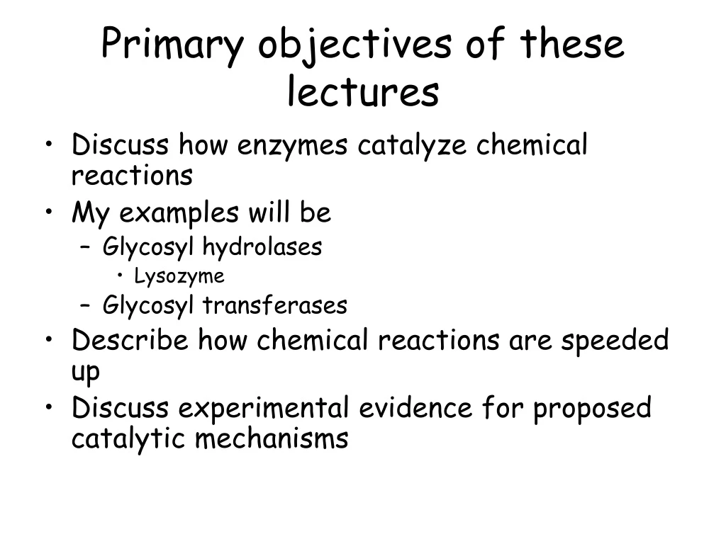
Primary objectives of these lectures • Discuss how enzymes catalyze chemical reactions • My examples will be • Glycosyl hydrolases • Lysozyme • Glycosyl transferases • Describe how chemical reactions are speeded up • Discuss experimental evidence for proposed catalytic mechanisms
Glycosidic bond cleavage H2O Glycone Aglycone • Example is lysozyme • Discovered by Alexander Fleming in 1920s • Something fell from his nose onto his bacterial agar plate • Bacteria lysed • Potential antimicrobial enzyme • He discovered a better antimicrobial agent later; what is it?
Glycosidic bond cleavage in free solution Glycone Aglycone H2O Transition state oxocarbenium ion attacked by hydroxyl ion
Rate of glycosidic bond cleavage • The transition state (positively charged oxocarbenium ion) is a very high energy molecule • Geometry changes from chair to half-chair • Why? • So C1 and ring oxygen are in same plane • So positive charge is not just at C1 but shared between C1 and ring oxygen • This stabilises positive charge. • Need lots of energy to cause change in geometry of sugar O5 C1
Two different mechanisms of acid-base assisted catalysis • Single displacement mechanism • Inversion of the anomeric configuration of glycone sugar β-glycosidic bond Bond is equatorial α-glycone sugar OH is axial
Two different mechanisms of acid-base assisted catalysis • Double displacement mechanism • Retention of the anomeric configuration of glycone sugar β-glycosidic bond Bond is equatorial β–glycone sugar: OH remains equatorial
Two different mechanisms of acid-base assisted catalysis • How does an enzyme generate protons and hydroxyl ions? • Two amino acids with carboxylic acid side-chains • Glutamate or aspartate • Two mechanisms are as follows:
Acid-base assisted single displacement mechanism Catalytic acid Catalytic base • The acid catalyst • Uncharged • Hydrogen in the perfect position to be donated to the glycosidic oxygen. • The catalytic base • Extracts a proton from water • Hydroxyl ion in the perfect position to attack C1 of the transition state
Acid-base assisted double displacement mechanism Catalytic acid-base Catalytic nucleophile • Two distinct reactions • Glycosylation • Formation of a covalent glycosyl-enzyme intermediate (ester bond) • The aglycone sugar released from active site • Deglycosylation • The ester bond between the glycone sugar and the enzyme is hydrolysed and the glycone sugar is released from the active site
Hen egg white lysozyme • The first enzyme structure solved • The textbook example of enzyme catalyzed glycoside hydrolysis • Hydrolyses the glycosidic bond via a retaining mechanism
Three possible mechanisms for lysozyme Covalent glycosyl-enzyme Asp 52 Ion-pair with long-lived glycosyl cation Covalent oxazoline (anchimeric assistance)
Accumulation of a 2F-chitobiosyl lysozyme (E35Q) intermediate 14315 Da GlcNAc2FGlcF Lysozyme (E35Q) 14864 Da Lysozyme (E35Q) 14316 Da Mass (Da) 2F-Chitobiosyl-lysozyme (E35Q) Free lysozyme
Stable covalent intermediate • Only the high molecular weight molecule observed • Suggests the formation of a covalent substrate-enzyme intermediate • How do we prove this? • Solve the 3D structure of this protein • Will show if there is a covalent bond between the substrate and enzyme
And the lysozyme mechanism is revisited: Covalent enzyme intermediate for hen egg white lysozyme Lysozyme (E35Q) Asp52 Vocadlo et al. Nature 412, 835-8
How can we identify the catalytic amino acids • Glycoside hydrolases are grouped in enzyme families based on sequence similarity (i.e. evolved from a common ancestor. Currently 100+ families • http://afmb.cnrs-mrs.fr/CAZY/ • All members of same family have • Evolved from the same progenitor sequence • Conserved mechanism • Same fold • Conserved catalytic apparatus
CAZY • Several families have ancient ancestral relationship • Same fold, mechanism and catalytic residues • How does CAZY help us? • Tells us what the catalytic residues are • Tells us the mechanism • Tells us the likely substrate specificity
Catalytic acid Sequence 1:73 QNGQTVHGHALVWHPSYQLPNWASDSNANFRQDFARHIDTVAAHFAGQVKSWDVVNEALFDSADDPDGRGSAN 1 UNIPROT:XYNA_PSEFL 1:73 335:407 QNGQTVHGHALVWHPSYQLPNWASDSNANFRQDFARHIDTVAAHFAGQVKSWDVVNEALFDSADDPDGRGSAN 2 UNIPROT:Q9AJR9 1:68 111:178 RHNQQVRGHNLCWHE--ELPTwaSEVngNAKEILIQHIQTVAGRYAGRIQSWDVVNEAILPKDGRPDG----- 3 UNIPROT:GUX_CELFI 3:66 115:176 --GKELYGHTLVWHS--QLPDWAKNLNGsfESAMVNHVTKVADHFEGKVASWDVVNEAFADG-DGP------- 4 UNIPROT:Q59277 3:61 116:173 --GKELYGHTLVWHS--QLPDWAKNLNGsfESAMVNHVTKVADHFEGKVASWDVVNEAFAD------------ 5 UNIPROT:Q59675 1:63 324:391 ENNMTVHGHALVWHSDYQVPnwAGSAE-DFLAALDTHITTIVDHYegNLVSWDVVNEAIDDNS---------- 6 UNIPROT:Q59301 2:63 343:409 -NNINVHGHALVWHSDYQVPNFmsGSAADFIAEVEDHVTQVVTHFkgNVVSWDVVNEAINDGS---------- 7 UNIPROT:Q59139 1:73 111:180 QNGKQVRGHTLAWHS--QQPGWMQssGSSLRQAMIDHINGVMAHYKGKIVQWDVVNEAFADG--NSGGRRDSN 8 UNIPROT:Q7SI98 1:73 73:142 QNGKQVRGHTLAWHS--QQPGWMQssGSTLRQAMIDHINGVMGHYKGKIAQWDVVNEAFSD--DGSGGRRDSN 9 UNIPROT:XYNB_THENE 1:62 96:158 KNDMIVHGHTLVWHN--QLPGWLTgsKEELLNILEDHVKTVVSHFRGRVKIWDVVNEAVSDS----------- 10 UNIPROT:Q60044 1:62 96:158 KNDMIVHGHTLVWHN--QLPGWLTgsKEELLNILEDHVKTVVSHFRGRVKIWDVVNEAVSDS----------- 11 UNIPROT:AAN16480 1:62 96:158 KNDMIVHGHTLVWHN--QLPGWLTgsKEELLNILEDHVKTVVSHFRGRVKIWDVVNEAVSDS----------- 12 UNIPROT:Q7TM36 8:68 2:58 -------GHTVVWHGA--VPTWLNasTDDFRAAFENHIRTVADHFRGKVLAWDVVNEAV---ADDGSG----- 13 UNIPROT:Q7WVV0 1:62 96:158 ENDMIVHGHTLVWHN--QLPGWITgtKEELLNVLEDHIKTVVSHFKGRVKIWDVVNEAVSDS----------- 14 UNIPROT:Q7WUM6 1:62 96:158 ENDMIVHGHTLVWHN--QLPGWITgtKEELLNVLEDHIKTVVSHFKGRVKIWDVVNEAVSDS----------- 15 UNIPROT:Q9WXS5 1:62 96:158 ENDMIVHGHTLVWHN--QLPGWITgtKEELLNVLEDHIKTVVSHFKGRVKIWDVVNEAVSDS----------- 16 UNIPROT:Q9P973 1:57 120:176 QNGKSIRGHTLIWHS--QLPAWVNnnNAdlRQVIRTHVSTVVGRYKGKIRAWDVVNE---------------- 17 UNIPROT:Q9X584 1:63 115:176 QNGKQVRGHTLAWHS--QQPGWMQssGSALRQAMIDHINGVMAHYKGKIAQWDVVNEAFADGS---------- 18 UNIPROT:XYNA_STRLI 1:63 114:175 QNGKQVRGHTLAWHS--QQPGWMQssGSALRQAMIDHINGVMAHYKGKIVQWDVVNEAFADGS---------- 19 UNIPROT:Q8CJQ1 1:63 114:175 QNGKQVRGHTLAWHS--QQPGWMQssGSALRQAMIDHINGVMAHYKGKIVQWDVVNEAFADGS---------- 20 UNIPROT:P79046 1:62 93:155 QNGQGLRCHTLIWYS--QLPGWVSSGNWN-RQTLEahIDNVMGHYKGQCYAWDVVNEAVDDN----------- 21 UNIPROT:Q9XDV5 3:71 427:505 --GMKVHGHTLVWHQ--QTPAWMndSGGNirEemRNHIRTVIEHFGDKVISWDVVNEAMSDNPSNpdWRGS-- 22 UNIPROT:Q8GJ37 3:71 427:505 --GMKVHGHTLVWHQ--QTPAWMndSGGNirEemRNHIRTVIEHFGDKVISWDVVNEAMSDNPSNpdWRGS-- 23 UNIPROT:Q7X2C9 1:63 27:88 QNGKQVRGHTLAWHS--QQPGWMQssGSSLRQAMIDHINGVMNHSKGKIAQWDVVNEAFADGS---------- 24 UNIPROT:Q9RJ91 3:61 105:162 --GMDVRGHTLVWHS--QLPSWVSPLGadLRTAMNAHINGLMGHYKGEIHSWDVVNEAFQD------------ 25 UNIPROT:Q59922 3:61 119:176 --GMKVRGHTLVWHS--QLPGWVSPLAadLRSAMNNHITQVMTHYKGKIHSWDVVNEAFQD------------ 26 UNIPROT:Q9RMM5 1:61 113:172 QNGKEVRGHTLAWHS--QQPYWMQssGSDLRQAMIDHINGVMNHYKGKIAQWDVVNEAFED------------ 27 UNIPROT:BAD02382 1:61 113:172 QNGKEVRGHTLAWHS--QQPYWMQssGSDLRQAMIDHINGVMNHYKGKIAQWDVVNEAFED------------
Why did the active centre of mannosidases look like that of glucosidases ?
Covalent Intermediate OS2 skew-boat Catalytic base Catalytic base Proves the identity of the catalytic nucleophile and shows sugar distortion Why is this remotely interesting?
Strong support for B2,5 transition-state • Enzymes perform mannoside chemistry by placing O2 pseudo-equatorial • The “difference” between mannosidases and glucosidases is actually seen at O3
Inhibitors of glycoside hydrolases • Glycoside hydrolase activities contribute to significant diseases • Flu • Type II diabetes • Possibly Cancer and Aids • To combat diseases need to develop inhibitors
Designing glycoside hydrolase inhibitors • What comprises a good inhibitor? • Mechanistic covalent inhibitors not used • Very high affinity non-covalent competitive inhibitors • Transition state inhibitors
glycosylation deglycosylation The retaining mechanism Transition state has a positive charged nature as leaving group departure precedes nucleophile attack
TS-based inhibitors that mimic charge distribution deoxynojirimycin Both have nM Ki values. Affinities are about one million times higher than substrate isofagamine Why are they transition state mimics? Contains a positive charge
Mimicking the half-chair • Insert a double-bond to enforce planarity
Drugs that mainly mimic the half chairAll picomolar affinities 108-fold tighter binders than substrates HIV drug: prevents glycosylation in mammalian cells AIDs virus surface proteins are not glycosylated and thus can’t evade the immune system Type II diabetes (inhibits human Amylase) Anti-flu drugs
Flu • RNA virus • Uses membrane of its host into which it inserts its own proteins • Major prevention of disease? • Vaccination • Pandemic • Killed 20 million 1918-199 • New pandemic about to occur. Bird flu coming from Far East China • Estimated 50000 will die in UK as a result of the new strain
Nucleotide-donor domain Acceptor domain Open conformation closed conformation Structure and function of glycosyltransferases that glycosylate macrolide antibiotics: Underpins the modulation of the specificity and potency of these antibiotics
Two folds • Both have two Rossman domains • GTA strongly linked may look like a single b-sheet • GT-B has two separate domains • Requirement of nucleotide binding limits number of folds greatly
Inverting GT Retaining GT
Inverting GT Retaining GT
References • Cantarel et al (2008) Nucleic Acid Res 37:D233-8 (CAZY) • Vocadlo at al. (2001) Nature 412:835-8. (Mechanistic inhibitors of glycoside hydrolases) • Lairson et al. (2008) Ann. Rev. Biochem. 77:521-555 (glycosyltransferases) • Rye and Withers (2000) Curr. Opin. Chem. Biol. 4:573-580 (glycoside hydrolases) • Tailford (2008) Nature Chem. Biol. Nat. 4:306-12 (Transition state geometry)