Download
1 / 59
640 likes | 1.27k Views
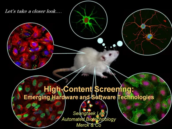
Fluorescent Imaging Solutions for High Content Screening. Herman Fennema Field Application Scientist Imaging and Microscopy April, 2006. Molecular Probes Campus, Eugene Oregon. HCS, HCA, HTI, AI, What are they?. High Content, Context Screening or Analysis (H.C.S.)

E N D
Fluorescent Imaging Solutions for High Content Screening Herman Fennema Field Application Scientist Imaging and Microscopy April, 2006
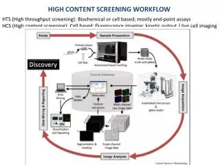
HCS, HCA, HTI, AI, What are they? • High Content, Context Screening or Analysis (H.C.S.) • High Content Analysis (H.C.A.) • High Throughput Imaging (H.T.I.), Automated Imaging (A.I.) • The one constant is that spatial information is derived. HCS Defined. First Proposed in 1986.
The C’s in HCS- Cell, Content, Context Less Searching More Content
Invitrogen HCS Product Overview • Enabling probes • HCS CellMask™ cytoplasmic/nuclear stains • Organelle markers • Assays • HCS LipidTOX™ phospholipidosis and steatosis kit • Apoptosis assays • Other cytotoxicity assays • GeneBLAzer™ assays • Expression tags • Tetracysteine and TC-FlAsH™ TC-ReAsH™ products • Antibodies • Alexa Fluor® and Qdot® conjugated antibodies
The C’s in HCS- Cell, Content, Context Less Searching More Content
Segmentation Tools- Customer Needs Segmentation is a Key Step in HCS Workflow
HCS CellMask ™ Stain- Red • Ex/Em 622/645 • ‘Tunable’ cytoplasmic/nuclear staining • Low conc. - comparable level of staining in cytoplasm and nucleus • High conc.- stronger staining in nucleus • Workflow compatibility • No wash needed- Last step in the staining and should be kept in the buffer during imaging 5 uM 1 uM Segmentation using 5 uM (on BD Pathway HT)
Why CellMask™ Stains Choices of HCS CellMask ™Stains- Multiplexing Flexibility
Intro to Cytotoxicity Cytotoxicity- Multi-Facet Phenomenon
Cytotoxicity Assays Wheel of Misfortune
HCS LipidTOX™ Kit • Phospholipidosis • Intracellular accumulation of phospholipids • Triggered by cationic amphiphilic drugs • Steatosis • Intracellular accumulation of neutral lipids • Triggered by drugs that affect metabolism of fatty acids and neutral lipids • HCS LipidTOX™ Kit • One workflow, two readouts • A complete assay kit for phospholipidosis and steatosis • With control compounds
Cytoskeleton: Tubulin • T34075 TubulinTracker™ Green • A11126 anti–α-tubulin mouse monoclonal IgG
Viability Assay Live Dead, Cytotoxicity Assay (L3224) In live cells, esterases convert Calcein-AM from non-fluorescent to green. Dead cells accumulate Ethidium Homodimer, the membrane impermeable nucleic acid stain to stain the nucleus red.
Endoplasmic Reticulum • E12353 ER-Tracker™ Blue-White DPX • E34250 ER-Tracker Red™ • E34251 ER-Tracker Green™ • S34200 SelectFX™ Alexa Fluor® 488 Endoplasmic Reticulum Labeling Kit • B7449 Brefeldin A, BODIPY® 558/568 conjugate *isomer 1*
Cytoskeleton: Actin • A12379 Alexa Fluor® 488 phalloidin • R415 Rhodamine phalloidin • A12381 Alexa Fluor® 594 phalloidin
Plasma Membrane • V22885 Vybrant® DiI cell-labeling solution • V22886 Vybrant® DiO cell-labeling solution • V22887 Vybrant® DiD cell-labeling solution • W11262 Alexa Fluor® 594 wheat germ agglutinin • F35355 FM® 1-43FX *fixable analog of FM® 1-43 membrane stain*
Live Cell Stains that are fixable (or retainable stains) The most Workflow friendly set of dyes!
Live Cell Stains and Live Cell-Fixable Stains Staining Alive, Staining Alive…
Live Cell Staining Solutions • Vybrant Assays for Live Cell Study • Vybrant® Phagocytosis Assay Kit • Vybrant® CFDA SE Cell Tracer Kit • Vybrant® MTT Cell Proliferation Assay Kit • Vybrant® Multidrug Resistance Assay Kit • Vybrant® Cell Adhesion Assay Kit (Calcein AM and SYTOX® Green) • Vybrant® Cell Lineage Tracing Kit – NEW! • Vybrant® Cell Metabolic Assay Kit *with C12-resazurin • Vybrant® Cytotoxicity Assay Kit *G6PD release assay – NEW! • Vybrant® Alexa Fluor® 488 or 555 or 594, Lipid Raft Labeling Kit • Vybrant® FAM Poly Caspases Assay Kit *for flow cytometry • Vybrant® Tubulin Staining Kit *for live cells* *100 assays – NEW!
Reagents for Multiple Apoptotic Parameters Timeframes within Camptothecin / Jurkat Model*
The C’s in HCS- Cell, Content, Context Less Searching More Content
More on Qdot® Nanocrystals CdS for UV-blue, CdSe for the bulk of the visible spectrum, CdTe for the far red and near-infrared. Qdots are bright: Extinction Coefficient Qdots (.5 to 2 x 10⁶ M-1 cm-1) Similar to RPE1 x 106. Organic dyes ~5-10 x 104. Quantum yields are high as well and they glow longer, so Qdots ~ 10 – 20x brighter. By 2-photon, their action cross section volume is 3 orders of magnitude higher. Excellent labels for conjugation. Brightness provides high sensitivity. Ideal for Multiplexing !
Qdot® Nanocrystals Optimal For: • Single line devices, with multiple detectors (7-8), FLOW, Imaging, uPlate. • Light Microscopy to Electron Microscopy (Ellisman, Deernick). • Two Photon, Small Animal In Vivo Imaging (SAIVI). • Confocal 3 D. • Arrays? When background comes down. • Live cell surface interrogation – primary conjugations. • Archival Pathology. • Single molecule detection. • Sensors? • FRET DONORS. • Multiplex RNAi – Lipofectamine 2000 + Dots + siRNA. Quantum Dot Corp. and BioPixel Staff in Eugene now
The C’s in HCS- Cell, Content, Context Less Searching More Content
Zymed® Research 1º Antibodies: 12 Categories • Angiogenesis • Apoptosis • Cancer Markers • Cell Adhesion, Cytoskeleton, & Matrix • Cell Junctions • Cell Cycle, Tumor Suppressors, & Oncogenic Proteins • DNA Replication & Repair; RNA Splicing & Translation • Neurobiology • Phospho-Amino Acid Antibodies • Signal Transduction (includes receptors, kinases, phospho-proteins) • Transcription Factors • Ubiquitin/Proteasome Protein Degradation Pathway Research portfolio focused on cancer, cell biology/signaling, and neuroscience
Multiplexing Alexa Fluor® Dyes across the spectrum Color Selection ♦ Brightness ♦ Photostability
Amplification and Multiplex FISH Multiplex detection of RNA expressed (7 genes) in Drosophila embryos FISH Tag RNA or DNA kits now optimized and available
The C’s in HCS- Cell, Content, Context Less Searching More Content
Stealth™ RNAi for target validation Stealth™ RNAi has improved specificity and stability
The C’s in HCS- Cell, Content, Context Less Searching More Content
Advantages of the GeneBLAzer®: platform • Robust: • Ratiometric readout reduces variation/ assay noise, e.g. due to cell number • Fast: • Rapid generation of stable cell lines by flow cytometry • Sensitive: • No mammalian background. Detection of <100 BLA molecules / cell • Miniaturizable: • Readily miniaturizable to 1536 & 3456 well formats An optimal detection system for assay development & HCS
The C’s in HCS- Cell, Content, Context Less Searching More Content
√ √ √ Why Expression Tags for In-Cell Labeling? Live Cell Compatible Protein Specific Labeling
Biarsenical labeling reagents bind to TC tags Tetracysteine (TC) tag is CCPGCC (adds ~1 kD) Figure from G. Parker TC tag = 6 amino acid tag
Tetracysteine tags are relatively small ReAsH Tetracysteine avGFP 505/528 nm 595/610 nm FlAsH-EDT2 ReAsH-EDT2 Green and Red fluorescent versions
Site-specific labeling in living cells CFP TC-FlAsH FRET shows specificity of labeling
Live cell imaging of mammalian cells Low background in un-transfected neighboring cells
Protein dynamics in living cells Monitor cytoskeletal changes or translocation events
TC-FlAsH™ II Data - Imaging/Bkg Control New wash protocol increase the S/N by almost 100%. Reduced Background by New Wash Protocol
Thank -You • Iain Johnson • Mike Janes • Yih Tai Chen • Ian Clements • Jason Kilgore • Dani Hill • Jerrod Salisbury • Kary Oakleaf • Mike O’Grady • Alex Savtchenko • Elena Molokanova • Jeffrey Dzubay • Jeff Hung • Beth Browne • Kathy Free • Brett Williams