
Prevalence of Osteoporosis in Mexico City Women
Study on low bone mineral density and osteoporosis in Mexico City's well-educated women. Factors impacting bone health and prevalence. Research uses data from private practice population.

Prevalence of Osteoporosis in Mexico City Women
E N D
Presentation Transcript
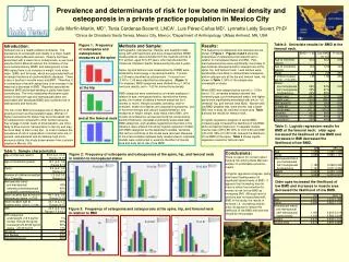
Prevalence and determinants of risk for low bone mineral density and osteoporosis in a private practice population in Mexico City Julio Morfín-Martín, MD1, Tania Cárdenas-Becerril, LNCA1, Luis Pérez-Cañas MD1, Lynnette Leidy Sievert, PhD2 1Clinica de Climaterio Santa Teresa, Mexico City, Mexico; 2Department of Anthropology, UMass Amherst, MA, USA Table 2: Univariate results for BMD at the femoral neck. Figure 1: Frequency of osteopenia and osteoporosis: measures at the spine Introduction: Osteoporosis is a health problem worldwide. The prevalence of overweight and obesity is a major health problem among Mexican women. Obesity has been associated with a lower risk of osteoporosis, so we could assume that in Mexican women the incidence of low bone mineral density (BMD) and osteoporosis is low. With aging there is an increase in weight, body mass index (BMI), and fat mass, which are associated with an increased incidence of cardiometabolic diseases. There is also a decline in muscle mass and BMD. Therefore, it is contradictory that with aging there is an increase in fat mass but a decrease in BMD. Reported associations between BMD and breast-feeding or parity have been contradictory. The only relationship that seems quite certain is that both age and menopausal status are associated with decreased BMD and increased risk of osteoporosis and fractures. The risk of low BMD and osteoporosis in Mexico is of growing concern as the population ages. Women of higher socioeconomic status may be at increased risk for osteoporosis compared to other women because they engage in lower levels of physical labor, are more likely to drive cars for transportation in the city and may be more likely to diet to stay thin. In order to assess the prevalence of risk in a population of women who are, in general, well-educated and of relatively high socio-economic status, this study draws women from a private practice in Mexico City. Methods and Sample: Demographic, reproductive, lifestyle, and treatment data, along with anthropometric and bone mineral density (BMD) measurements, were collected from the medical records of 511 women, aged 30 to 87 years, who had attended the Clinica de Climaterio Santa Teresa during the past 5 years. Spine, hip and femoral neck measurements of BMD were obtained by dual-energy x-ray absorptiometry. T-scores <-2.50 were classified as osteoporosis. T-scores from -2.49 to -1.00 were classified as osteopenia. (Figure 1) For analyses, BMD categories were divided into <-1.00 for low bone density, and >-1.00 for normal bone density. BMD categories were examined by univariate analyses in relation to age, menopausal status, reproductive history (parity, the number of babies a woman breast fed for 4 months or more), lifestyle variables (smoking, level of exercise), health (hot flashes and depressive symptoms), use of hormone therapy and calcium supplementation, and measures of height, weight, body mass index (BMI), arm muscle circumference, and percent body fat (computed by skinfold thickness). Variables significantly associated with BMD categories, and variables regarded as important in the literature, were entered into binary logistic regression models with BMD categories as the dependent variable. Variables that did not contribute to the model were removed. Because of the intercorrelation between body measurements, separate models were constructed to examine the effect of muscle area and body fat on risk of low BMD. Results: The frequency of osteopenia and osteoporosis are shown in Figure 1. Figures 2 and 3 show the frequencies of osteopenia and osteoporosis in relation to menopausal status and BMI. Post-menopausal women were significantly more likely to demonstrate osteopenia and/or osteoporosis at the spine, hip, and femoral neck. Lean women were significantly more likely to demonstrate osteopenia and/or osteoporosis at the hip and femoral neck. As shown in Table 1, 50% of the sample was overweight or obese. When BMD was categorized as normal (>-1.00)or low (≤-1.0), univariate analyses showed that increasing age, post-menopausal status, and higher parity were significantly associated with low (≤-1.0) vertebral, hip, and femoral neck BMD. Women with low BMD weighed less, were shorter, had a lower BMI, less % body fat, and less muscle area.Table 2.shows the results for femoral neck. In logistic regression analysis on spinal BMD, increasing age increased the likelihood of low BMD, while increasing height and, in separate models, muscle mass (OR 0.98, 95% CI 0.97-0.99) and BMI (OR 0.92, 95% CI 0.87-0.96) reduced the likelihood of low BMD at the spine. Table 3 shows logistic regression results for femoral neck. at the hip and at the femoral neck P<0.001 Table 3: Logistic regression results for BMD at the femoral neck: older ages increased the likelihood of low BMD and increases in BMI decreased the likelihood of low BMD. Table 1: Sample characteristics Conclusions: There is reason for concern about fracture risk among these Mexican women of comfortable economic means. In logistic regression analyses, parity and breast feeding were not significant determinants of BMD. It appears that increasing muscle mass is almost as protective for women at risk for low BMD as increasing BMI. Although level of exercise was not associated with BMD in this study, the results of exercise, i.e., increasing muscle area, do appear to reduce the likelihood of low BMD and exercise should be encouraged. Figure 2: Frequency of osteopenia and osteoporosis at the spine, hip, and femoral neck in relation to menopausal status P<0.05 P<0.001 P=0.04 P<0.001 Older ages increased the likelihood of low BMD and increases in muscle area decreased the likelihood of low BMD. Figure 3: Frequency of osteopenia and osteoporosis at the spine, hip, and femoral neck in relation to BMI P<0.001 P<0.01 ns Sunset in the city of Campeche.


