Download

1 / 36
360 likes | 547 Views
On the menu at top click on “Slide Show” and then click on “From Beginning” , If this opens in PowerPoint, otherwise just click to start. Southwestern Illinois EMS System. Introduction to Cardiac Anatomy and Physiology. Introduction. Cardiovascular disorder

E N D
On the menu at top click on “Slide Show” and then click on “From Beginning” , If this opens in PowerPoint, otherwise just click to start.
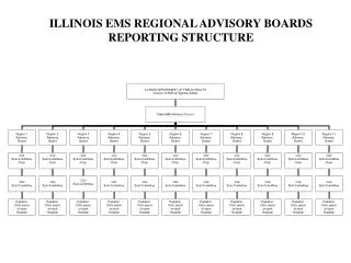
Southwestern Illinois EMS System Introduction to Cardiac Anatomy and Physiology
Introduction • Cardiovascular disorder • Diseases, conditions that involve heart, blood vessels • Heart disease • Conditions affecting heart
Introduction • Coronary heart disease • Coronary arteries, resulting complications • Angina pectoris, acute MI • Coronary artery disease • Affects arteries that supply heart muscle with blood
Risk Factors & Prevention Strategies • Risk factors • Nonmodifiable (fixed) risk factors • Modifiable risk factors • High blood pressure • Elevated serum cholesterol levels • Tobacco use • Diabetes
Risk Factors & Prevention Strategies • Risk factors • Modifiable risk factors • Physical inactivity • Obesity, body fat distribution • Metabolic syndrome
Risk Factors & Prevention Strategies • Risk factors • Contributing risk factors • Stress • Inflammatory markers • Psychosocial factors • Alcohol intake
Anatomy Review • Blood vessels • Arteries • Arterioles • Capillaries • Venules • Veins
Anatomy Review • Heart anatomy • Location • Mediastinum • Behind sternum, above diaphragm • Base • Apex
Anatomy Review Heart Location
Anatomy Review • Heart anatomy • Heart chambers • Upper chambers • Right, left atria • Lower chambers • Right, left ventricles
Anatomy Review • Heart anatomy • Septum • Pulmonary circulation • Systemic circulation • Blood carried from heart to body through arteries, arterioles, capillaries • Blood returned to heart through venules, veins
Anatomy Review Heart anatomy • Heart layers • Endocardium • Myocardium • Epicardium • Pericardium
Anatomy Review • Heart anatomy • Heart valves • AV valves • Separate atria from ventricles • Tricuspid valve, between right atrium, right ventricle • Mitral/bicuspid valve lies between left atrium, left ventricle • Open when forward pressure forces blood forward • Close when backward pressure pushes blood backward
Anatomy Review • Heart anatomy • Atrial kick • Blood flows continuously into atria • 70% flows directly through, into ventricles before atria contract • When atria contract, additional 30% added to filling of ventricles • When ventricles contract (systole), pressure rises • Tricuspid, mitral valves close when pressure within ventricles exceeds that of atria
Anatomy Review • Heart anatomy • Semilunar (SL) valves • Pulmonic, aortic valves • Prevent backflow of blood from aorta, pulmonary arteries into ventricles • Close as ventricular contraction ends, pressure in pulmonary artery, aorta exceeds that of ventricles • Chordaetendinae, connective tissue, attached to AV valves underside & papillary muscles
Anatomy Review • Blood flow through heart • Enters right atrium via superior, inferior venaecavae, coronary sinus • Right atrium through tricuspid valve into right ventricle • Right ventricle expels blood through pulmonic valve into pulmonary trunk • Flows through pulmonary arteries to lungs
Anatomy Review • Blood flow through heart • Low in O2, passes through pulmonary capillaries • From left atrium through mitral valve into left ventricle • Distributed throughout body through aorta, its branches
Anatomy Review • Blood flow through heart • Tissues of head, neck, upper extremities via superior vena cava • Lower body via inferior vena cava • Superior, inferior vena cava carry contents into right atrium
Anatomy Review • Cardiac cycle • Repetitive pumping process, events associated with blood flow through heart • Systole • Diastole
Anatomy Review • Cardiac cycle • Depends on cardiac muscle ability to contract, condition of heart’s conduction system • Pressure with each chamber rises in systole, falls in diastole • Conduction system provides timing of events between atrial, ventricular systole
Anatomy Review • Coronary arteries • Right, left • Main arteries • Left anterior descending (LAD), left circumflex (LCX), right coronary artery (RCA) • Lie on outer surface of heart
Anatomy Review Coronary Arteries
Anatomy Review • Coronary veins • Travel alongside arteries • Coronary sinus, largest vein, drains heart
Anatomy Review • Heart rate • Affected by sympathetic, parasympathetic ANS • Chronotropic effect • Inotropic effect • Dromotropic effect
Anatomy Review • Heart rate • Baroreceptors • Specialized nerve tissue (sensors) • Found in internal carotid arteries, aortic arch • Detect changes in blood pressure • When stimulated cause sympathetic/parasympathetic response • Will “reset” to new “normal” after few days of exposure to specific pressure
Anatomy Review • Heart rate • Chemoreceptors • In internal carotid arteries, aortic arch, medulla detect changes in concentration of hydrogen ions (pH), O2, carbon dioxide in blood
Anatomy Review • Heart rate • Parasympathetic stimulation • Parasympathetic fibers supply sinoatrial node, atrial muscle, & AV junction of heart by vagus nerves
Anatomy Review • Heart rate • Sympathetic stimulation • Sympathetic nerves supply specific areas of heart’s electrical system, atrial muscle, ventricular myocardium • When stimulated, norepinephrine released • Increases in heart rate shorten all phases of cardiac cycle
Anatomy Review • Heart rate • Increases in heart rate shorten all phases of cardiac cycle • Electrolyte, hormone levels, medications, stress, anxiety, fear, body temperature can influence heart rate • Heart rate increases when body temperature increases, decreases when body temperature decreases
Heart as Pump • Venous return • Most important factor determining amount of blood pumped out by heart is amount of blood flowing into right heart
Heart as Pump • Cardiac output • Amount of blood pumped into the aorta each minute by heart • Defined as stroke volume x heart rate • Stroke volume determined by • Preload • Afterload
Heart as Pump • Cardiac output • Frank–Starling’s law • Greater the volume of blood in heart during diastole (preload), the more forceful cardiac contraction & more blood ventricle will pump (stroke volume) • Important that heart adjust its pumping capacity in response to changes in venous return • During exercise, heart muscle fibers stretch in response to increased volume (preload) before contracting
Heart as Pump • Cardiac output • Frank–Starling’s law • Factors that increase cardiac output include increased body metabolism, exercise, age & size of body • Factors that may decrease cardiac output include shock, hypovolemia, heart failure
Conclusion Please complete the 10 question online exam and submit when completed. Thank You