OPHTHALMOSCOPY
350 likes | 763 Views
Learn general principles, normal & disease fundus changes. Master ophthalmoscope usage & exam techniques. Ideal for medical students.

OPHTHALMOSCOPY
E N D
Presentation Transcript
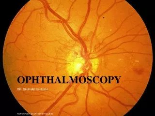
OPHTHALMOSCOPY DR. SHAHAB SHAIKH POWERPOINT COURTESY DR MUJEEB
OBJECTIVES • To explain the general principles of ophthalmoscopy • Describe the normal appearance of the fundus. • Describe the changes in the fundus that occur commonly in disease. • Appreciate the importance of performing ophthalmoscopy as a part of the routine physical examination.
METHOD • For a good view of fundus the pupil should be dilated by instilling few drops of short acting mydriatic drug (e.g.1% cyclopentolate). • The subject should be examined in sitting or lying down position. • Examination room should be dark. • keep the eye as still as possible.
Position of the examiner For examining right eye of the patient, • Examiner should stand on right side of the patient. • Hold the instrument in his right hand. • Use examiner’s right eye. If examining left eye, stand on left side, hold instrument in left hand use left eye.
Viewing should begin about half meter away from the eye. • First see the “Red reflex” • Initially the lens power in the instrument should be set to zero, or refractive error of patient or examiner, e.g. if the patient is myopic then set the (-ve )lens, if the examiner or patient is hypermetropic then set the lens to (+ve) lens. If both patient & examiner have refractive error then sum together their powers. • e.g. if examiner having +2, & pt. having +1 lens then adjust +3 lens in ophthalmoscope. • If examiner have +2 diopters lens & pt. having -4 diopters lens then adjust (+2)+(-4) =(-2) lens in ophthalmoscope.
DIABETIC RETINOPATHY On examination we find • Capillary micro-aneurysms are seen as tiny spots near the retinal vessels. • Retinal haemorrhages and exudate: • Hemorrhage appear round • Hard exudate (yellow with irregular margin) • New vessel formation
HYPERTENSIVE RETINOPATHY On examination we find • Generalized narrowing of retinal arteries. • Arterio venous nipping i.e. indentation of the veins when they are crossed by the arteries. • Retinal haemorrhages and exudate: • Flame shaped hemorrhages • Soft exudate (cotton wool) • Papilloedema.
Papilloedema Edema of optic nerve head, most commonly due to increased intracranial pressure. eg. Brain tumor. On examination of fundus we find; • Increased redness of disc with blurring of its margins. • Physiological cup disappears. • Retinal vessels are distended.
NORMAL OPTIC CUP DEEP OPTIC CUP