Download
1 / 28
280 likes | 407 Views
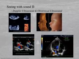
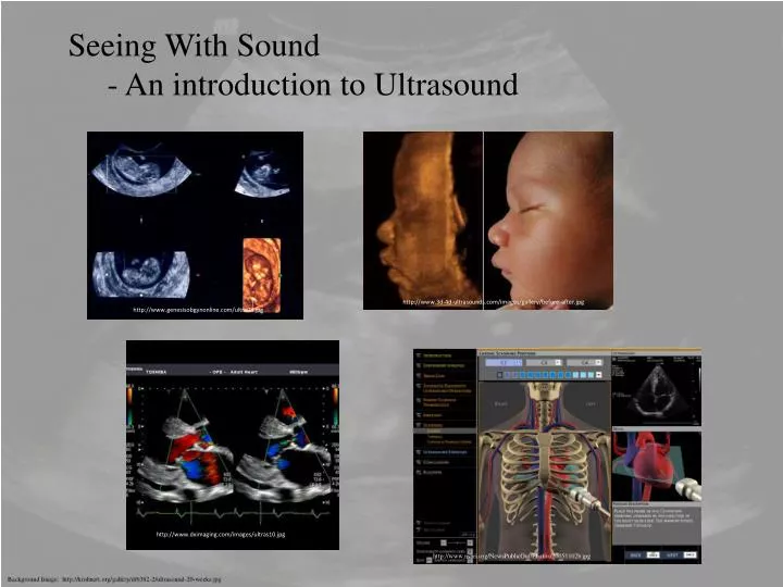
Seeing With Sound - An introduction to Ultrasound. http://www.genesisobgynonline.com/ultra08.jpg. http://www.3d-4d-ultrasounds.com/images/gallery/before-after.jpg. http://www.nsbri.org/NewsPublicOut/Photos/20051102b.jpg. http://www.dximaging.com/images/ultras10.jpg.

E N D
Seeing With Sound - An introduction to Ultrasound http://www.genesisobgynonline.com/ultra08.jpg http://www.3d-4d-ultrasounds.com/images/gallery/before-after.jpg http://www.nsbri.org/NewsPublicOut/Photos/20051102b.jpg http://www.dximaging.com/images/ultras10.jpg Background Image: http://krohnert.org/gallery/d/6382-2/ultrasound-20-weeks.jpg
Seeing With Sound - Outline • Introduction to Sound and sound waves. • The velocity of sound in materials. • Basics of ultrasound. • Homogeneous and non-homogeneous materials. • Reflections & echoes. • Intensity of ultrasound – the decibel scale. • Attenuation of ultrasound in homogeneous media.
Seeing With Sound - The Physics of Ultrasound: background • Most imaging technologies are concerned with the interaction of ionizing radiation (x-rays, g-rays, or particles) or electromagnetic interactions with tissues in the body. • Ultrasound is different – it is not an electromagnetic wave and it is non-ionizing. Sound is a mechanical wave – it needs a medium in order to propagate. • Ultrasound is high-frequency sound waves in the 1 – 10MHz range. • Ultrasound waves are traveling waves and not standing waves. • The energy associated with the waves can be absorbed by the material and the ultrasound waves can be reflected or refracted at different tissue boundaries. • Ultrasound image formation is accomplished by using echoes, or reflections from different media, such as organs, vessels or other structures. • Refraction produces distorted images and absorption is of little clinical value
Seeing With Sound - The Physics of Ultrasound: background • Sound is a longitudinal wave – medium oscillates parallel to the direction of propagation of the wave. There are regions of compression (higher than ambient pressure) and decompressions (lower than ambient pressure). • The human ear is able to hear sound in the frequency range of about 20 – 20,000 Hz. • Ultrasound refers to frequencies that are higher than 20-kHz (and for comparison with light, infrasound refers to frequencies that are lower than 20-Hz.) • The wave speed of a sound wave is related its frequency and the wavelength through: • For any material, the velocity of sound is nearly constant, meaning that the velocity is independent of frequency. Or there is little dispersion of the waves. • The wavelength of a sound wave is the physical distance between any two compressions or decompressions (rarefactions.) http://www.physics.uc.edu/~sitko/CollegePhysicsIII/14-Sound/Sound.htm
Seeing With Sound - The Physics of Ultrasound: background • Animation of a continuously vibrating source in a column of air perhaps, or sound propagation in a material. • Notice that the molecules do not move with the passing wave. Waves do not transport matter, but rather energy from one location to another. Application courtesy of Dan Russell, Kettering University • For our ultrasound applications we’ll actually be using a pulsed source of sound. This is what we’ll be calling a pulse-echo ultrasound. • This is how bats and submarines hunt their prey.
Seeing With Sound - The Physics of Ultrasound: background • To determine the speed of sound in a medium we choose to model the medium as a collection of atoms connected by springs and apply Newton’s laws of motion it is found that the speed of sound depends further on its density, r and it compressibility, K, through • The compressibility is defined as the fractional change in volume of material per unit increase in pressure • Thus a large compressibility means that it is easier to squeeze something.
Seeing With Sound - The Physics of Ultrasound : background • Ultrasound imaging is particularly useful in studying soft tissues that are not able to be contrasted with say using x-rays. • Doppler ultrasound can detect and monitor the flow of fluids, such as blood flow in arteries and veins. • Ultrasound is used (almost extensively) in obstetric and gynecologic, cardiac, vascular and abdominal imaging. • Ultrasound equipment is relatively cheap and portable, unlike and MRI or X-ray equipment. • Ultrasound does not produce ionizing radiation in the body. • Believed that the risks of using ultrasound as a diagnostic tool are low.
Seeing With Sound - The Physics of Ultrasound : an example • The fundamental frequency of a piano’s middle C key is 261.6 Hz. What is its wavelength in air? What is its wavelength in tissue? • What is the wavelength of a 1 MHz ultrasound signal in soft tissue? • What frequency ultrasound would be needed to image features that are 0.5mm in diameter?
Seeing With Sound - The Physics of Ultrasound • Sound waves are periodic mechanical disturbances that need a medium to propagate through. • Sound waves are longitudinal waves made up of repeating cycles of compression and decompression. • Disturbances due to sound radiate outward radially from the source causing disturbances in the local surrounding media. For example, a hammer hitting a nail. • Ultrasound makes use out of the reflections of high frequency sound waves. • The orientation of the ultrasound beam at the time of the pulse gives a bearing, or heading for the beam, toward a target. • The echoes that are received back yield distances to the target, if the return time of the echo and velocity of sound in the medium is known. • This process is known as SONAR (Sound Navigation And Ranging.)
Seeing With Sound - The Physics of Ultrasound • The basics of ultrasound are to send out a well defined, high frequency pulse of sound energy in a known direction, from a source (a transducer) and to detect the resultant fainter echoes. • The echoes are turned into an electrical signal (a voltage) and the voltages are processed and displayed on an oscilloscope (or TV screen.) • The transducer is constructed of a piezoelectric material. • A piezoelectric is a material that has a well defined oscillation frequency and the material is put into oscillation by an oscillating electric field (or by an oscillating current.) • The piezoelectric material can not only generate • but also detect sound vibrations. • Planar vs. other geometries – maybe • a curved surface piezoelectric would be • better? http://www.ndt-ed.org/EducationResources/CommunityCollege/Ultrasonics/EquipmentTrans/radiatedfields.htm
Seeing With Sound - The Physics of Ultrasound http://www.genesis.net.au/~ajs/projects/medical_physics/graphics/transducer.jpg • The transducer head contains the piezoelectric material and the transducer is acoustically coupled to the body by using a gel. • The gel minimizes reflections of the ultrasound at the skins surface and a beam of ultrasound pulses are delivered to the body. • The velocity of sound in soft tissue is approximately 1540-m/s. • The ultrasound pulse has an intensity I that decreases exponentially as it passes through the body’s tissues and organs. http://www.ndt-ed.org/EducationResources/CommunityCollege/Ultrasonics/EquipmentTrans/radiatedfields.htm
Seeing With Sound - The Physics of Ultrasound - Echoes • Material types: • Homogeneous medium – the medium is the same throughout – like a fluid filled bladder. • Medically these are uninteresting materials since fluid in the bladder will produce no echoes. • Non-homogeneous medium – the medium has a sharp boundary and the boundary will produce an echo. • Medically more interesting and the boundary could be different tissue densities or different tissue elasticities.
Seeing With Sound - The Physics of Ultrasound - Echoes • The time lapse from transmission of pulse to detection of echo is proportional to the depth within the tissue. This is called an A-type scan. A-type scans are used to measure distances. The transducer emits an ultrasonic pulse and the time taken for the pulse to be reflected and return to the transducer is graphed in order to determine how far away the object is. A-type scans only give one-dimensional information and therefore are not useful for imaging. There are several other scan types that we’ll look at. B-Type & Doppler http://www.genesis.net.au/~ajs/projects/medical_physics/ultrasound/index.html • The intensity of the echo signal increases with the amount of physical difference between the two tissues. Compare for example a tennis ball thrown off of a wall that is made out of brick to one made out of a blue tarp.
Seeing With Sound - The Physics of Ultrasound - Intensity • The intensity of sound is described by the decibel scale. • The threshold intensity is that which the ear is just barely sensitive to: Ithreshold ~ 1x10-12 W/m2. • The intensity ratio for two sounds as heard by your ear is given by: . • For ultrasound, the threshold intensity is the US beam intensity, IUS and the echo has an intensity, Iecho. • Thus we have . • Alternatively and coincidentally more useful, we could write this in terms of the ratio of the intensities . • It two signals differ in intensity by 20dB, what is the ratio of their intensities?
Seeing with Sound Ultrasound – Intensity • The attenuation, or loss of sound energy, is due to frictional forces at the atomic/ molecular level as the atoms/molecules vibrate and return to their relaxed state. • The intensity of the ultrasound is proportional to the square of the amplitude of the wave so that • Solving for the intensity ratio we have • Suppose that an echo is ½ as intense as the original US pulse, what is the intensity drop in decibels?
Seeing With Sound - The Physics of Ultrasound – Intensity • So how strong an echo will be produced? • It depends on many factors. • If the beam passes through a homogeneous medium like a fluid filled cyst or a large blood vessel, the ultrasound pulse simply propagates through and its intensity will simply decrease exponentially with increasing distance, x. • ms is called the ultrasound intensity attenuation coefficient in units of distance-1. • ms is a combination of the energy loss due to absorption by the tissue (and turned into heat) and energy loss due to scattering of the beam by irregularities. • If we take the base 10 logarithm of the equation above, (and then multiply by 10) we get
Seeing With Sound - The Physics of Ultrasound – Attenuation of US • The left hand side of this equation is the definition of the dB. Thus we have • And now we define a more useful quantity, the decibels of intensity loss (in dB per cm) as • The absorption coefficients are frequency dependant and for the most part that frequency dependence is linear in frequency. • To find the decibels of intensity loss (per cm) we use a graph (or a computerized data file which is programmed on an actual US machine) to determine the absorption coefficient at a particular frequency in a particular tissue. • Then the number of decibels of intensity loss is easily calculated and this is easier than calculating actual intensities.
Seeing With Sound - The Physics of Ultrasound – Attenuation of US • The attenuation given is for a one way travel. • As the ultrasound reflects back it is further attenuated. • Suppose that a 5 MHz ultrasound source is used to image a muscle, how many decibels of intensity are lost as the ultrasound passes one way through 2cm of muscle? • How about through 2cm of blood? Wolbarst, Physics of Radiology, Ch. 11
Seeing With Sound - The Physics of Ultrasound – Reflection & Refraction • Just like light, whenever sound energy passes from one medium in which the speed of sound is v1 to another medium in which the speed of sound is v2, the waves refract. • Using the geometry of the system, the law of refraction for sound is given as: • Here, like light, the frequency is constant across the interface so that the wave passes continuously from one medium to the other. • Thus we have a relation between the velocity and wavelength in the two media: • Refraction causes distortions in the images causing displacements in the image of objects in the scan and also cause some loss in resolution.
Seeing With Sound - The Physics of Ultrasound – Reflection & Refraction Wolbarst, Physics of Radiology, Ch. 11 Figure demonstrating the effects of refraction for sound as well as for light.
Seeing With Sound - The Physics of Ultrasound – Reflection at Acoustic Media Interfaces • So, let’s return to the question of how strong an echo will be? • For a homogeneous medium – a fluid filled cyst or a large blood vessel, US propagates and is attenuated but no echoes are formed. • If US propagates through dissimilar tissues or organs then multiple reflections are possible and energy is reflected back towards the transducer. • The return time is proportional to the displacement. • The intensity of the echo depend on this distance since it will be attenuated exponentially with distance but also on the difference in physical properties of the materials (density or elasticity.) • Sharp echoes are produced form a large flat boundary between materials that have significant physical differences. • The reflected waves obey the law of reflection.
Seeing With Sound - The Physics of Ultrasound – Reflection at Acoustic Media Interfaces • The elasticity of a material along with its density determine the speed of sound in the medium. • Thus where the velocity of sound changes abruptly is where the strongest echoes will be produced. • This means that more intense US waves will be reflected. • However, if two materials have similar elastic properties then the US waves will produce small reflections and most of the US will propagate into the second medium. • The amount of reflection of sound at an interface is determined by the acoustic impedance, Z, of the materials. • And using the definition of the velocity of sound , where the units are 1 kg/m2s = 1 Rayl.
Seeing With Sound - The Physics of Ultrasound – Reflection at Acoustic Media Interfaces • Why the acoustic impedance? Analogous to the an inverse index of refraction • Thus we can write the reflection coefficient in terms of the acoustic impedance. • And the transmission coefficient: T = 1 – R • Since Z is proportional to v, the greater the difference in Z’s the larger the reflection coefficient, R. • How much of an US wave is reflected at a muscle-fat interface? How much is transmitted? (Zmuscle = 1.7x10⁶ kg/m2s ; Zfat = 1.38x10⁶ kg/m2s) • Compare to muscle-bone or muscle-air. (Zair =400kg/m2s; Zbone= 5x10⁶ kg/m2s)
Seeing With Sound - The Physics of Ultrasound – B or brightness-type Scans • A –or amplitude-type scans give a one dimensional view of an object. We’d like more spatial information. • To do this we would still like distance information, but we’d also like to see the variation in US intensity from place to place. • To do this we sweep the transducer across an area of the body producing what’s called a B-type scan. • A B-type scan is a combination of many A-type scans that are displayed on a computer or TV screen.
Seeing With Sound - The Physics of Ultrasound – B or brightness-type Scans • A single pulse of ultrasound passing into a series of tissues will give rise to a series of spots, with the brightness of the spots corresponding to the amplitude of the reflection from different layers. • The largest amplitude gives rise to a spot with the greatest brightness corresponds to white. • The smallest amplitude gives rise to a black spot. • Intermediate amplitudes give various shades • of gray. • The area that does not give rise to any spike • for example the aqueous and the • vitreous will appear black http://brighamrad.harvard.edu/Cases/bwh/hcache/96/findings.html http://www.mrcophth.com/commonultrasoundcases/principlesofultrasound.html#B%20in US image of a gallstone lodged in the cystic duct.
Seeing With Sound - The Physics of Ultrasound – Advantages of US • Ultrasound examinations are non-invasive i.e. they do not require the body to be opened up, or anything to be inserted into the body. This is a major advantage compared to fiber optic endoscopy, for example, which may involve much more patient discomfort as the probe is inserted. • Ultrasound methods are relatively inexpensive, quick and convenient, compared to techniques such as X-rays or MRI scans. The equipment can be made portable, and the images can be stored electronically. • No harmful effects have been detected, at the intensity levels used for examinations and imaging. This contrasts with methods based on X-rays or on radioactive isotopes, which have known risks associated with them, and ultrasound methods are preferred whenever possible. This is particularly relevant to examination of expectant mothers. • Ultrasound is particularly suited to imaging soft tissues such as the eye, heart and other internal organs, and examining blood vessels.
Seeing With Sound - The Physics of Ultrasound – Disadvantages of US • The major disadvantage is that the resolution of images is often limited. This is being overcome as time passes, but there are still many situations where X-rays produce a much higher resolution. • Ultrasound is reflected very strongly on passing from tissue to gas, or vice versa. This means that ultrasound cannot be used for examinations of areas of the body containing gas, such as the lung and the digestive system. • Ultrasound also does not pass well through bone, so that the method is of limited use in diagnosing fractures. It is possible to obtain quite good ultrasound scans of the brain, but much greater detail is obtained by an MRI scan.
For Wednesday, September 30, 2009: Read Kane Chapter 4, sections 4.5 – 4.8 Wolbarst, Chapter 11, sections 11.1 – 11.12 and do Kane, Chapter 4 Questions: Q4.3, Q4.4, & Q4.5 & Problems: P4.1, P4.2, & P4.3 For Wednesday, I’ll collect these Q’s and P’s as well as the three due last Friday, Questions Q3.1 & Q3.5 & Problem Q3.5. For Friday, October 2, 2009 Read Kane Chapter 4, sections 4.8 – 4.11 and do Kane Chapter 4, Question: Q4.2 & Problems: P4.5, 4.6, & 4.7 For Monday, October 5, 2009 Read Kane Chapter 4, sections 4.12 – 4.18 and do Kane Chapter 4, Question Q4.6, & Problems: P4.12 & P4.14 For Wednesday, October 7, 2009 I will collect the following: Kane Chapter 4, Questions Q4.2 & Q4.6 and Problems P4.5, P4.6, P4.7, P4.12, & P4.14