Download
1 / 17
180 likes | 457 Views
Interstitial Lung Disease (ILD) Mike McFarlane (CT1) 12/5/12. SLIME. What we’ll cover. Definition Different types of ILD Pathophysiology Presentation Investigations Management Prognosis Clinical Scenario Summary. What we won’t. Other causes of restrictive lung defects

E N D
Interstitial Lung Disease (ILD)Mike McFarlane (CT1)12/5/12 SLIME
What we’ll cover • Definition • Different types of ILD • Pathophysiology • Presentation • Investigations • Management • Prognosis • Clinical Scenario • Summary
What we won’t... • Other causes of restrictive lung defects • Thoracic cage defects • Anything fun!!
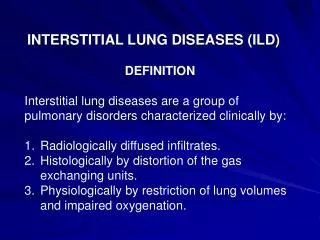
Definition • Interstitial lung disease is a diverse group of conditions affecting the pulmonary interstitium and/or the alveolar lumen • Not airways! • The gas exchange parts and support tissue
Different types of ILD • Idiopathic Pulmonary Fibrosis AKA: Cryptogenic fibrosingalveolitis, Usual interstitial pneumonitis • Extrinsic Allergic Alveolitis AKA: Hypersensitivity pneumonitis • Other Industrial lung diseases • Sarcoidosis
Pathophysiology • Cycles of inflammation, scarring and fibrosis leads to irreversible damage to interstitial tissue • IPF: ?autoimmune • EAA: • Farmer’s Lung: Micropolysporafaeni/ AspergillusFumigatus • Bird Fancier’s Lung: Avian serum proteins • Malt worker’s Lung: Aspergillusclavatus • Inhalation (humidifier) fever: Thermophillicactinomycetes • Other “dust” diseases: (CABS) • Coal Workers Pneumoconiosis: Coal Dust • Caplan’s Syndrome: CWP + Rheumatoid arthritis • Asbestosis: Asbestos • Beryllosis: Beryllium • Silicosis: Silicon • Sarcoidosis: Multisystem autoimmune non-caseatinggranulomatous disorder
Presentation • Symptoms • SOB on exertion • Lethargy • Dry cough • EAA – variability with work; IPF – progressive worsening • Sarcoid • extrapulmonary features: • Anterior uveitis, conjunctivitis, arthralgia, erythemanodosum • SMOKING, OCCUPATION, PETS • Signs • Tachypnoeic • Clubbing – IPF, EAA • Cyanosis • Fine end-inspiratorycreps • IPF – more at bases • EAA – more at apices
Investigations • Bedside • PEF , including work and home measurements • Sats, RR • Blood tests • FBC, U&Es, LFTs, CRP, ESR • ABG - hypoxia • ANA and RF can be + in IPF • Calcium can be high in Sarcoidosis (serum ACE can be high but not always!) • Imaging • CXR – fine reticulonodular shadowing • CT • Ground glass appearance • Honey combing
Investigations (2) • Special tests • Spirometry • Restrictive defect i.e. Lung volume reduced. FVC reduced, FEV1 reduced in proportion (or slightly less). Therefore FEV1:FVC is normal or high!! • TLCO (transfer factor) reduced – due to fibrosis of alveolar walls. Means a thicker barrier to gas exchange and less effective transfer • N.B. Bronchoscopy, BAL, Biopsy
Management • Conservative • Smoking cessation • Change working conditions • Change work • Medical • Depends on cause!! • IPF – very little besides oxygen! Steroids of little help • EAA – Steroids for acute episodes • Sarcoidosis – Only treat if extrapulmonary manifestations! Steroids • Surgical • Lung transplant
Prognosis • IPF – poor • EAA – depends on extent of disease and ability to avoid the cause • Industrial lung disease - variable • Sarcoidosis - variable
Clinical scenario • A 64 year old gentleman presents to his GP with increasing SOB over the last 6 months. • His exercise tolerance has reduced to the point where walking to the corner shop makes him out of breath. • He also complains of a dry cough. • He has a past medical history of high blood pressure which is managed with Ramipril. • He has never smoked and works as an office manager • He has no pets • On examination he is slightly short of breath with O2 sats 93% on air and he has clubbing. Auscultation reveals bilateral basal fine end inspiratorycrepitations and no wheeze.
Clinical scenario Cont • What are your main differentials for this gentleman? • How would you investigate this gentleman? • What is your management plan? • Will anything help?
Summary • ILD is more complicated than it needs to be • Cardinal features are: • Fine end inspiratorycreps– due to fibrosis • The cause of the fibrosis will usually be hinted at in occupation or hobbies! • If no cause obvious its probably IPF • Restrictive spirometrywith reduced gas transfer • Treatment depends on cause – usual steroids