Download
1 / 32
330 likes | 390 Views
Explore the transformative potential of ultrasound technology for paramedics, enhancing diagnostic accuracy, treatment precision, and patient outcomes. Embrace the shift towards ultrasound as a primary diagnostic tool in EMS, revolutionizing healthcare practices.

E N D
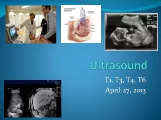
Prehospital Ultrasound “The future stethoscope” Peter Bonadonna, EMT-P CI/C June 2011 Disclosure: No financial or other benefit is provided to me, my family or my place of work for advancing and promoting this material at this time.
Presentation Goals • To introduce the practicing paramedic to a technology that will revolutionize the way we examine and treat our patients in the near future. • To excite you about the utility of ultrasound. • To give a short introduction about the numerous medical and trauma ultrasound uses in EMS. • To dispel fear and misinformation that is holding this technology back. • To leave you supporting this technology.
What is ultrasound? The use of high frequency sound waves (> 20 kHz) to accomplish a task such as echolocation or US imaging These frequencies are far above the human hearing range. 1-20 MHz is typical medical ultrasound range
Therapeutic UltrasoundThis is not what we are going to talk about Ultrasound energy is applied to generate heat energy to promote muscle relaxation and improved circulation.
Now you can take a look inside. Prehosptial Focused Assessment with Sononography in Trauma. It will definitely change your care. Paramedics tend to be very visual learners. This is perfect for learning and using Ultrasound. A picture is worth 1000 words.
Dynamic assessments See the size, shape and function in real time !
So Peter, “Why are you so interested in advancing field ultrasonography?” • I see once-valued EMS treatments are now deemed questionable or injurious (Trendelenburg, MAST, ET, IV fluids, some medications). The value of the old style paramedic is in peril. • I have been observing many misses and wrong Dxs by good providers who relied on physical exam. • I have noticed declining physician confidence in paramedics. • I recognize that this technology is a real game-changer for paramedics. It would empower us to be far more accurate, give better care and shift the paradigm from educated guessing and treatment to accurate diagnosis and accurate treatment.
How can ultrasound help Paramedics? • Educationally • Students can see actual anatomy and real-time function on classmates and themselves. It provides another reason to really learn anatomy well. • Students can appreciate the variability of human anatomy. • It’s fun, which helps in the learning and retention process. • Even if they work where no US is available students will be better providers. • Clinically • Can see inside the body, far more accurate diagnosis and trending. • More exacting treatments are possible and increased documentation. • Better triage of our patients to Aeromedical, ED, CT or the OR. • Professionally • A chance for the Paramedic profession to elevate its abilities and accuracies. • Better patient care will immediately be recognized by the medical community as well as by the public. • Better self-esteem when accuracy improves.
Why now ? • The price has dropped substantially and the technology has reached the point of “will be smaller, cheaper and clearer from here on in”. • Physio/Sonosite collaboration – Your next heart monitor may have ultrasound built in. • Several leading physicians are promoting EMT-P’s use ultrasound but most paramedics are oblivious (or resistant) to the notion. • Initial entry-level exams, like P-FAST, are easy to learn, easy to do, accurate, noninvasive, feasible, rapid, versatile, repeatable, time saving and very safe. • Better for patients (early accurate Dxs = more timely care).
Why now ? Cont. • Takes less time to perform than a 12 lead EKG and can be done in transit (unlike 12 Lead). Easier to teach how to “read”. • US can significantly increase diagnostic accuracy and therefore treatment accuracy. • US can increase skills success – ET, IV, needle thoracostomy, pericardiocentesis, external pacing. • The need for accuracy in diagnosis and treatment is greater than ever before i.e. for physician trust & the medicolegal climate of today. • Serial ultrasounds may be as valuable to the Physician as serial 12 lead ECGs have been. • Because if we don’t do it, someone else will.
You have had many of these cases. How often did you make the correct call ? Not suspected – actually called it in as these ? • AAA or ruptured AAA • Thoracic bleeding (hemothorax) • Pericardial effusion from cancer, uremia, heart failure, s/p cardiac procedure or trauma • Intraperitoneal bleeding • Ectopic pregnancy or ruptured ectopic pregnancy • Pneumothorax • Cardiogenic shock from low EF (pump failure) • Obstructive uropathy (kidney stone, bladder distention, Foley obstruction) • Pleural effusions as the cause of SOB These aren’t rare. They’re just always missed by EMS
The physical exam fallacy • So you think that history and physical exam will make you pretty accurate in your assessments? • AAA (>50% missed on physical exam by Drs) • Cardiac Tamponade (Impossible to Dx w/o imaging) • TNT (Recent studies show many EMS misDx) • Shock (fluid or pressors, if you guess wrong pt. is harmed) • Internal Bleeding (can have normal VS until too late) • OB (paramedics have never been able to really examine) • SOB Cardiac Asthma vs. Asthma/COPD. • NTG, CPAP, diuretics vs. β2 , Hydration
The reasons it has not taken off • So new that you may have not heard about it. • You may have already prejudged it. • Someone you respect has told you its bad. * • “Most EMT-Ps haven’t mastered 12 leads and you want them to do this?!?!” Remember this is easier! • “Show me the supporting studies.” • Still pictures of US can be very hard to interpret and scare people away. The dynamic image is much easer to read! The probe location is known and you can look around. • Fear of having to really make a diagnosis. • Fear of having to learn more. It’s not a lot. I promise.
Prehospital Focused Assessment with Sonography in Trauma or “P-FAST” Paracolic gutters Literally “next to the colon”. A potential space where blood or fluid naturally trickles into. This is why we only have to look in three places with the probe to detect bleeding. Detects as little as 250 mls of blood in the abdomen and 20 mls in the chest.
Examples • Intraperitoneal Bleeding
Morrison’s Pouch(Hepatorenal pouch) Significant intraperitoneal bleeding Normal
Pneumothorax Negative predictive value for pneumothorax 100%
Examples Lung in M-mode
28 yo ♂with history of blunt trauma to the left chest • Pt is SOB, has diminished breath sounds on the left and has a sat of 90% P 90 BP 130/95 R 18 • Point tenderness over several ribs 5-7 on Lt. • 10 minutes into transport patient becomes acutely and severely short of breath. P120 R 32 BP 80/p Sat. 80% Breath sounds hard to hear. • Distance to the hospital 20 minutes at best • What would most paramedics do? This patient does nothave a pneumo or tension pneumothorax and you can now quickly hunt for the cause of their distress with your ultrasound machine and exam. This would prevent a paramedic from ever needling a normal chest. Right now this is a 1 in 4 occurrence.
49 yo ♀with hx of asthma Mild bilateral wheezing. VS BP 158/90 P 80, RR 30, SpO2 96%, NSR, 12 lead normal, Treatment ? This patient has heart failure and can be made worse by beta agonists which strengthen the RV more than the left increasing pulmonary congestion. A normal ventricle ejects 50-60 percent of the blood out of the chamber. This ejection fraction (EF) is about 10 percent. RV LV LA After treatment with albuteral she says she feels worse!
Does the patient need fluid? 60 yo M. Vomiting, SOB, P 90 BP 130/80 R 20 normal skin turgor Orthostatic Vital Signs can not be done on all patients and when they can be performed, are not always reliable Click here to see how you can answer this question
Looking at the IVC is important Hypovolemia, sepsis, neuro Heart failure/ pulmonary embolus, cardiac tamponade
Cardiac Arrest Are you treating all of your arrests the same way? i.e. Standard ACLS? Do you wonder why the overall survival has not changed in 25 years? There are many different causes of cardiac arrest in addition to arrhythmia. This requires specific treatment strategies that could be delivered in the field where it counts the most.
Incremental training and use After we master and become comfortable with the simple P-FAST Exam we can move on to: • Aorta Assessments (identify AAA or dissection) • ET assessment (can immediately see tube in esophagus) • FEER- Focused Echo Evaluation in Resuscitation / PE/ Pacing • Pregnancy – IUP, Fetal HR, Age, position, Ectopic • IV starts • Ocular (Retinal Detachment, ONSD – elevated ICP) • Lung Ultrasound (easily differentiate CHF vs. COPD) • Kidney/Gall stones • Adult Epiglottitis, Long Bone Fracture, Surgical airway
Are there any EMS systems currently doing Ultrasound? • Over 20 agencies in the USA. >200 devices • Boston EMS • Austin and Odessa, Texas • Winnemucca, Nevada • Temple Terrace, Florida • California • HCMC EMS, Minneapolis, Minnesota • Norfolk, Virginia
What are the short-comings? Brand new, high end machines are expensive. Some of the smaller machines are delicate and have less functionality. No reimbursement at this time. Some patients can’t be fully imaged due to body size or rarely subcutaneous emphysema. It is operator dependent. (but so is physical exam) Not all EDs are up to speed on this so they may be resistant.
Are any Paramedic Programs teaching it? • Monroe Community College Paramedic Program, Rochester, NY • ProEMS Paramedic College, Cambridge, Mass.
Should all paramedics use this? I believe the answer should be No. We need to introduce it carefully into each system. Only highly motivated, knowledgeable paramedics should be chosen at first. High volume or experienced providers first. Only trained and credentialed providers (can be local EMS or hospital based credentials). Only those who participate in USCME. Only those with medical director approval.
Thank you for your attention For More Information Visit: www. ParamedicUltrasound.com Contact Peter for Intro session like this one or for training in P-FAST 6 and 24 hour courses