Hypoperfusion and Shock
Hypoperfusion and Shock. Hypoperfusion. Common problem Extent makes resuscitation difficult Shock due to hypoperfusion Start fluid resuscitation as soon as possible. Overview. Describe differences between compensated and uncompensated shock

Hypoperfusion and Shock
E N D
Presentation Transcript
Hypoperfusion • Common problem • Extent makes resuscitation difficult • Shock due to hypoperfusion • Start fluid resuscitation as soon as possible
Overview • Describe differences between compensated and uncompensated shock • Review differences of distributive, non-distributive and obstructive shock • Explore pathophysiology for different etiologies of shock • Discuss interventions for early and late shock
Physiology BP = Cardiac Output x Systemic Resistance Cardiac Output = Stroke Volume x Heart Rate After-load = Resistance to blood being ejected Pre-load = Blood returned to heart Starling’s Law = Amount of cardiac muscle stretch LifeART NHTSA
Children Increased heart rate Vasoconstriction Prolonged compensation Rapid decompensation Adults Increased stroke volume Vasoconstriction Tachycardia Slow, but sustained compensation Shock Compensation Children vs. Adults EPS411.com
Categories of ShockNon-Distributive Hypovolemic • Hemorrhagic • Metabolic
Categories of ShockDistributive • Anaphylaxis • Septic • Neurogenic
Categories of ShockObstructive • Pulmonary embolus • Tension pneumothorax • Cardiac tamponade
Etiologies of Hypoperfusion(Common) • Emesis and diarrhea • Osmotic diuresis from diabetes • Internal or external blood loss • Plasma loss from sepsis or anaphylaxis
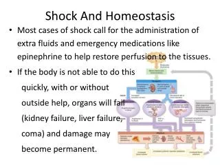
Etiologies of Hypoperfusion(Uncommon) • Spinal cord injury • Cardiac failure Medications required to restore perfusion
Severity of HypoperfusionCompensated Compensated Decompensated Signs are due to inadequate tissue perfusion Volume Compensated shock is reversible with fluids Time
Severity of HypoperfusionCompensated Shock Signs Compensated Decompensated Volume Pulse Breathing Blood Pressure AVPU Altered Mental Status Time
Severity of HypoperfusionCompensated Shock Signs Compensated Compensated Decompensated Decompensated Weak or absent peripheral pulses, weak central pulses Volume Weak peripheral pulses, strong central pulses EPS411.com Time
Severity of HypoperfusionDehydration Testing Hypovolemic patient’s skin will “tent”
Severity of HypoperfusionDecompensated Shock Compensated Decompensated Inadequate tissue perfusion to all organs Volume Body is unable to continue compensation Time
Severity of HypoperfusionDecompensated Shock Signs V P U Compensated Decompensated Volume Pulse Breathing Blood Pressure AVPU Altered Mental Status Weak or absent peripheral pulses, weak central pulses Time
Scene Survey Hazards to you, your partner, the patient and bystanders
First ImpressionPediatric Assessment Triangle Compensated or decompensated EPS411.com
First ImpressionGeneral Appearance • Observe interactions • Not sick - attentive to environment, focus on familiar people and objects, alert for threats • Good brain function requires adequate oxygenation, ventilation, cerebral perfusion • Sick - does not care you are present or recognize parents
First ImpressionGeneral Appearance • Muscle tone • Spontaneous movements • Skin color • Other signs of distress KyleDavidBates.com
First ImpressionCirculation to the Skin Skin color, capillary refill, distal vs. central pulses
First Impression Yes No Sick Not Sick Significant MOI? Rapid Initial Assessment Appropriate Interventions Relationship Transport Priority Involve Family Transport Method Detailed History Transport Destination Focused Physical Exam
Initial AssessmentAirway Loss of airway may occur in decompensated shock Identify and treat life threats
Initial AssessmentBreathing Assess for chest trauma Abnormal sounds Rate effort and volume Administer O2 and treat cause
Initial AssessmentCirculation Compensated Weak peripheral pulses, strong central pulses Decompensated Weak or absent peripheral pulses, weak central pulses EPS411.com EPS411.com
Initial AssessmentCirculation Management – Intravenous • Fluid bolus if any signs of shock • Early recognition of hypoperfusion and fluid resuscitation are key • Select a large bore catheter • Location close to central circulation • Two IVs may be needed
Initial AssessmentCirculation Management – Intraosseous Can be used on any age child
AnatomyNeonate Leg Cross Section Skin Intraosseous Catheter Tibia Subcutaneous Fat Lateral Compartment Anterior Compartment Fibula Posterior Compartment
Other IssuesIO Insertion • Depth based on patient size and weight • Gently insert catheter • Advance catheter slowly • Feel needle drop into medullary space • Frequently monitor insertion site and extremity • Need hands-on training
IO Insertion Anatomical Landmarks Tibial Tuberosity Medial Tibia Patella
IO Insertion Unable to Palpate Tibial Tuberosity Finger Width Finger Width Often difficult or impossible to palpate
IO Insertion Able to Palpate Tibial Tuberoisty Finger Width
AnatomyNeonate Leg Cross Section Traditional IO Catheter Tibia Fibula Left Leg
Anatomy11 y.o. Tibia Cross Section Insertion Site Tibia Fibula Left Leg
Initial AssessmentCirculation Management – Crystalloids 20 mL/kg, < 20 minutes Reassess patient after each fluid bolus
Initial AssessmentNever Administer D5W D5W can lead to hyperglycemia
Initial AssessmentCirculation Management – Medications Sepsis Pressers and antibiotics Cardiogenic Shock Pressers, furosemide, morphine and antiarrhythmics Anaphylaxis Epinephrine, diphenhydramine, Solu-Medrol EPS411.com
Initial AssessmentCirculation Management – Medications Use medications after fluid boluses EPS411.com
Transport Decision Rapid transport for pediatric shock patients
Focused HistoryQuestions to Determine Type of Shock Bleeding Vomiting Diarrhea Fluid intake / urine output Fever Anaphylaxis signs FEMA Photo Library / Andrea Boomer
Summary • Recognition and rapid intervention are keys to treatment • Pulse quality and level of consciousness are key indicators • Obtain IV or IO access if shock treatment is needed • Deliver crystalloid fluids at 20 mL/kg