Download
1 / 27
270 likes | 461 Views
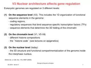
V3 Nuclear architecture affects gene regulation. Eukaryotic genomes are regulated on 3 different levels: On the sequence level (V2). This includes the 1D organization of functional sequence elements in the genome: - coding regions,

E N D
V3 Nuclear architecture affects gene regulation • Eukaryotic genomes are regulated on 3 different levels: • On the sequence level (V2). This includes the 1D organization of functional sequence elements in the genome: • - coding regions, • - regulatory sequences that bind sequence-specific transcription factors (TFs) • - sequence elements that determine the 3D folding of the chromatin • On the chromatin level (V1, V5-V8) • - different histone compositions • - the ¨histone code¨ (see lectures on epigenetics) • On the nuclear level (today) • - the 3D structure and functional compartimentalization of the genome inside the interphase nucleus. Driel et al. J. Cell. Sci. 116, 4067 (2003) Biological Sequence Analysis
Nuclear architecture A striking feature of nuclear architecture is the existence of distinct structural and functional compartments. Well characterized nuclear substructures include the nuclear lamina, nucleoli, PML and Cajal bodies, and nuclear speckles. Also, a growing number of components of the machinery that is required for transcription or its repression are known to have a non-homogeneous distribution in the nucleoplasm. At the level of the genome itself, the genetic material is folded and packaged in the nucleus into higher-order structures that are likely to contribute to the regulation of gene expression. Lanctot et al. Nat. Rev. Genet. 8, 104 (2007) SS 2009 – lecture 3 Biological Sequence Analysis 2
Organization of the mammalian cell nucleus The nucleus is characterized by a compartmentalized distribution of functional components. The nuclear envelope contains pores and rests on a meshwork of intermediate filaments, the nuclear lamina. Nucleolar organizer regions cluster to form nucleoli. In the chromosome territory–interchromatin compartment (CT–IC) model, chromatin is organized in distinct CTs. Also depicted are nuclear speckles, PML bodies and Cajal bodies located in wider IC lacunas. Major goal: identify the principles that govern the spatial organization of the genome. Lanctot et al. Nat. Rev. Genet. 8, 104 (2007) SS 2009 – lecture 3 Biological Sequence Analysis 3
Model of functional nuclear architecture (a) CTs have complex folded surfaces. Inset: topological model of gene regulation. A giant chromatin loop with several active genes (red) expands from the CT surface into the IC space. Cremer, Cremer Nat. Rev. Gen. 2, 292 (2001) SS 2009 – lecture 3 Biological Sequence Analysis 4
Molecular model of nuclear pore complex Localization volumes of all 456 proteins in the NPC (excluding the FG-repeat regions) in 4 different views. The proteins are colour-coded according to their assignment to the 6 NPC modules. Alber et al. Nature (2007)
Chromatin movement In vivo studies showed that the positions of labelled chromatin are constrained during interphase within a radius of ca. 0.5 – 1 m. This is less than 1% of the volume of a typical spherical mammalian nucleus that has a diameter of 10 m. Only during early G1, long-range movements of 2 m are observed. In Drosophila, labelled topoisomerase II that binds to a heterochromatic repeat block on chromosome X could explore about half of the radius of a Drosphophila nucleus (2 m) indicating constrained diffusion. Current work addresses whether the chromatin movements are correlated with changes in gene expression. Lanctot et al. Nat. Rev. Genet. 8, 104 (2007) SS 2009 – lecture 3 Biological Sequence Analysis 6
Nuclear architecture affects gene regulation One classical example of organization within the nucleus is the distinction between decondensed, transcriptionally active euchromatin and more condensed, generally inactive heterochromatin. Individual chromosomes occupy distinct positions in the nucleus, referred to as chromosome territories. As a result of different compaction levels, different chromosome segments adopt a complex organization and topography within their chromosome territory. Gene-rich regions tend to be oriented towards the nuclear interior, whereas gene-poor regions tend to be oriented towards the periphery. This principle of nuclear organization is evolutionarily conserved. Lanctot et al. Nat. Rev. Genet. 8, 104 (2007) SS 2009 – lecture 3 Biological Sequence Analysis 7
Chromosome territories in the chicken (a-c) staining of different chromosomes. (d) optical section through a chicken fibroblast nucleus showing various mutually exclusive CTs. Cremer, Cremer Nat. Rev. Gen. 2, 292 (2001) SS 2009 – lecture 3 Biological Sequence Analysis 8
The interchromatin compartment LASER CONFOCAL sections through a HeLa cell nucleus with GFP-tagged H2B, and staining of speckles a Section showing GFP-tagged chromatin (high density, white; low density, grey), two nucleoli (nu) and the interchromatin compartment (IC) space (black). Note the variability in the width of this space with examples of IC lacunas (asterisks). Inset: expansions of less condensed chromatin into the IC space at higher magnification. b Speckles visualized in the same section using antibodies to the non-snRNP splicing factor SC-35. c Overlay of sections (chromatin, green; speckles, red) shows that speckles form clusters in IC lacunas. These lacunas are only partially filled by the speckles, leaving space for other non-chromatin domains. … … Cremer, Cremer Nat. Rev. Gen. 2, 292 (2001) SS 2009 – lecture 3 Biological Sequence Analysis 9
Regulation on the nuclear level (1) A classical example is the nucleolus, in which the rRNA-coding gene clusters of several chromosomes are brought together to create a subnuclear domain that is dedicated to rRNA synthesis and processing and pre-ribosome synthesis. (2) Clustering of heterochromatin e.g. near the nuclear envelope. Difficulty: we lack experimental tools to manipulate nuclear structure! So far, researchers have mostly analyzed correlations. Driel et al. J. Cell. Sci. 116, 4067 (2003) SS 2009 – lecture 3 Biological Sequence Analysis 10
Model of structural constraints on chromatin mobility A model of structural constraints on chromatin mobility and gene– gene interactions. a Three hypothetical chromosome territories — green, blue and red — are shown. Lanctot et al. Nat. Rev. Genet. 8, 104 (2007) SS 2009 – lecture 3 Biological Sequence Analysis 11
Model of structural constraints on chromatin mobility Filled circles: Subchromosomal ~1 Mb domains. For the green territory, the lighter-green circles indicate gene-poor chromatin; for all territories, dark-coloured circles indicate gene-dense chromatin. Dark background shading: areas that contain transcriptionally active genes. As the chromatin moves over time, there are concomitant changes in the positioning of genes that are involved in interchromosomal interactions (the positions of 3 such genes, one from each territory, are indicated by black circles around the ~1 Mb domains in which they are located). b | The three panels show how the chromatin that is located where the same three territories are adjacent to each other (shown as a shaded region in panel a) might become repositioned over time; the panels indicate 3 consecutive time points during interphase. Lanctot et al. Nat. Rev. Genet. 8, 104 (2007) SS 2009 – lecture 3 Biological Sequence Analysis 12
The multiloop subcompartment model b, c Two 3D models of the internal ultrastructure of a ~1-Mb chromatin domain. b The nucleosome chain is compacted into a 30-nm chromatin fibre and folded into ten 100-kb-sized loop domains according to the multiloop subcompartment model. Occasionally, 30-nm fibres are interrupted by short regions of individual nucleosomes (small white dots). The arrow points to a red sphere, with a diameter of 30 nm, that represents a TF complex. Cremer, Cremer Nat. Rev. Gen. 2, 292 (2001) SS 2009 – lecture 3 Biological Sequence Analysis 13
The multiloop subcompartment model b, c Two 3D models of the internal ultrastructure of a ~1-Mb chromatin domain. c | Each of the ten 100-kb chromatin domains was modelled under the assumption of a restricted random walk (zig-zag) nucleosome chain. Each dot represents an individual nucleosome. Nine 100-kb chromatin domains are shown in a closed configuration and one in an open chromatin configuration with a relaxed chain structure that expands at the periphery of the 1-Mb domain. The open domain will have enhanced accessibility to partial transcription complexes preformed in the interchromatin compartment. By contrast, most of the chromatin in the nine closed domains remains inaccessible to larger factor complexes (arrows). Cremer, Cremer Nat. Rev. Gen. 2, 292 (2001) SS 2009 – lecture 3 Biological Sequence Analysis 14
Localization at the nuclear envelope • Eukaryotic genomes contain 3 classes of chromatin. The establishment and maintenance of chromatin states is related to their spatial distribution with the interphase nucleus. • Open or actively transcribed chromatin, which contains genes with engaged RNA polymerases. • Potentially active chromatin, which contains promoters that are poised to respond to activating signals, but from which stable transcripts are rare or non-existent. • In yeast, these two states account for the vast majority of chromatin. • (3) In mammals, they only comprise a small fraction of the genome. In differentiated somatic cells, most DNA is in a transcriptionally silent heterochromatic state. Here, genes are generally repressed, gene promoters are inaccessible to TFs. Akhtar, Gasser, Nat. Rev. Genet. 8, 507 (2007) SS 2009 – lecture 3 Biological Sequence Analysis 15
Transcriptionally silent heterochromatic states Positioning of chromatin at the nuclear envelope can contribute to gene regulation in both a positive and negative manner. Sites that anchor silent chromatin are mechanistically distinct from those for active genes (budding yeast experiments). In budding yeast, heterochromatin binds the nuclear envelope through Esc1 (green) which forms distinct foci with nuclear pores (Nup49 labelled red). Akhtar, Gasser, Nat. Rev. Genet. 8, 507 (2007) SS 2009 – lecture 3 Biological Sequence Analysis 16
Localization to nuclear envelope In yeast nuclei, envelope-associated proteins such as Esc1 (enhancer of silent chromatin 1) are present in foci at the peripheri. However, they do not coincide with the pores (Immuno-EM). Esc1 binds Sir4 (silent information regulator 4) which is an integral component of repressed heterochromatin in yeast. This interaction is necessary and sufficient to anchor silent chromatin at the nuclear envelope. Akhtar, Gasser, Nat. Rev. Genet. 8, 507 (2007) SS 2009 – lecture 3 Biological Sequence Analysis 17
Localization to nuclear envelope In metazoan nuclei, the nuclear envelope is underlaid by a continuous meshwork of lamins and lamin-associated proteins (LAPs) which preferentially associate with inactive chromatin regions. Akhtar, Gasser, Nat. Rev. Genet. 8, 507 (2007) SS 2009 – lecture 3 Biological Sequence Analysis 18
Putative role of NPC in coupling transcription and mRNA processing by gene looping in yeast. Although not all transcriptional activity in the nucleus will be subject to this mode of regulation, the budding yeast NPC seems to work together with transcriptional activation mechanisms to fine-tune gene activity. The SAGA chromatin-remodelling complex in yeast contains Sus1; Sus1 is also present in the mRNA-export complex TREX, which interacts with Nup1. Nup2 also interacts with the promoters of active genes, and the NPC-associated protein Mlp1 (myosin-like protein 1) accumulates at the 3′ end of active genes, where it contributes to an RNA surveillance mechanism. Optimal activation can require both localization of the induced gene at the NPC as well as at the 3′ UTR. Our model suggests that gene looping, which results from the coincident NPC-tethering of an initiation complex and mRNA-processing complexes that are associated with the 3′ UTR, will help to fine-tune the expression of certain genes. Finally, the pore protein Nup2 was found to tether genes through a histone variant H2A.Z (Htz1) in yeast. This could reflect a heritable localization that contributes to forms of epigenetic control. Akhtar, Gasser, Nat. Rev. Genet. 8, 507 (2007) SS 2009 – lecture 3 Biological Sequence Analysis 19
3C method: analyze gene interactions in 3D 3C stands for chromosome conformation capture method. Method measures the formation of crosslinks between chromatin segments after formaldehyde fixation of whole cells or isolated nuclei. A frequency of crosslinking above control levels indicates spatial proximity 4C method: combine 3C with microarrays. 3C and 4C methods indicate that long-range chromatin interactions (gene kissing) are involved in the epigenetic regulation of gene expression. One important example: H19 and Igf2 genes. Lanctot et al. Nat. Rev. Genet. 8, 104 (2007) SS 2009 – lecture 3 Biological Sequence Analysis 20
Regulatory models at imprinted loci (A) The enhancer–blocker model (also known as the boundary model) is well studied at the Igf2/H19 locus and consists of an imprinting control region (ICR) located between a pair of reciprocally expressed genes that controls access to shared enhancer elements. On the paternal allele, the differentially methylated domain (DMD) acquires methylation (black circles) during spermatogenesis, which leads to repression of the H19 promoter. The hypomethylated maternal DMD acts as an insulator element, mediated through binding sites for the methylation-sensitive boundary factor CTCF (shaded ellipse). When CTCF is bound, Igf2 promoter access to the enhancers (E) distal to H19 is blocked. Blue boxes : paternally expressed alleles, red boxes : maternally expressed alleles, black boxes : silenced alleles, grey boxes : nonimprinted genes. Arrows on boxes indicate transcriptional orientation. PLOS Genet. 2, e147 (2006)
Protein Interactions and Chromatin Loops • reading the imprint: candidate "imprinting transcription factors" CTCF, YY1 • chromatin loop model • DMRs interact via proteins • mediates interaction with the enhancers H19 Igf2 • Murrell et al. (2004) Nature Genet. 36: 889 • maternal chromosome: DMR1 and DMR unmethylated, CTFC bound H19 is expressed (interaction with the enhancers), Igf2 is silenced • paternal chromosome: DMR and DMR2 methylated, no CTCF binding Igf2 in contact with enhancers, active; H19 silenced
Gene kissing In this example of gene kissing, copies of the Drosophila melanogaster Fab7 regulatory element that are present on two different chromosomes co-localize in the cell nucleus. DAPI (4′,6-diamidino-2-phenylindole) is the DNA counterstain, sd shows the position of a transgenic Fab7 copy that is inserted in the X chromosome at the scalloped (sd) locus. Abd-B indicates the locus that is regulated by the endogenous copy of the Fab7 element. The two loci ‘kiss’ each other in a significant fraction of the nuclei, as seen in the merged panel. Lanctot et al. Nat. Rev. Genet. 8, 104 (2007) SS 2009 – lecture 3 Biological Sequence Analysis 23
How do compartments form? Two non-exclusive models have been proposed to explain how compartments form and are maintained. Compartments could correspond to pre-existing structures, or they could be formed as a result of self-assembly. It was reported in 1999 that maintenance of chromosome-territories requires RNA. For the case of the clustering of Fab7 transgenes, the presence of small 21-23 nt long RNA generated by the RNAi machinery was correlated with spatial co-localization. Mutations in the RNAi machinery disrupted the long-range Fab7 interactions. So also RNAs seem to have structural and regulatory roles in nuclear architecture. Lanctot et al. Nat. Rev. Genet. 8, 104 (2007) SS 2009 – lecture 3 Biological Sequence Analysis 24
Chromatin mobility in nucleus Chromatin mobility allows dynamic interactions between genomic loci and between loci and other nuclear structures. a and b show two chromosome territories. Within each territory a gene locus is indicated in red. Movement of chromatin is depicted by arrows. Two possible configurations are represented for each territory, with the dotted outline of one superimposed on the other. The transition involves repositioning of the two loci within the three-dimensional space of the nucleus. In this hypothetical example, the bottom configuration in each case is favored on transcriptional activation of the loci. Two alternative models have been proposed to account for the compartmentalization of nuclear functions that are involved in gene expression. Lanctot et al. Nat. Rev. Genet. 8, 104 (2007) SS 2009 – lecture 3 Biological Sequence Analysis 25
Chromatin mobility in nucleus a, compartments are pre-existing structures containing molecular machineries that are dedicated to specific nuclear functions. Movement of chromatin from one compartment to another leads to changes in expression of the corresponding genomic regions. Activation is triggered by repositioning of the gene loci to an activating compartment, away from silencing compartments. Lanctot et al. Nat. Rev. Genet. 8, 104 (2007) SS 2009 – lecture 3 Biological Sequence Analysis 26
Chromatin mobility in nucleus In b, compartments are transient self-organizing entities. In this case, gene activation leads to dissolution of the silencing compartments, changes in gene positioning and de novo assembly of an activating compartment. Once initiated, this state can be maintained by the self-assembly of components that are involved in gene regulation, as well as the clustering of chromatin regions that contain actively expressed genes. Lanctot et al. Nat. Rev. Genet. 8, 104 (2007) SS 2009 – lecture 3 Biological Sequence Analysis 27