Download
1 / 66
660 likes | 758 Views
Proteins, Enzymes, Biochemistry Sept. 21, 2001. Duncan MacCannel: Historical Perspective on Molecular Biology / Genetics. Background The Thread of Life. Susan Aldridge. Chapter 2 Molecular Biology of the Cell. Alberts et al. Garland Press Suggested further reading

E N D
Proteins, Enzymes, Biochemistry Sept. 21, 2001 Duncan MacCannel: Historical Perspective on Molecular Biology / Genetics
Background • The Thread of Life. Susan Aldridge. Chapter 2 • Molecular Biology of the Cell. Alberts et al. Garland Press • Suggested further reading • Protein molecules as computational elements in living cells. D. Bray. Nature. 1995 Jul 27;376(6538):307-12. • Signaling complexes: biophysical constraints on intracellular communication. D. Bray. Annu Rev Biophys Biomol Struct. 1998;27:59-75. • Metabolic modeling of microbial strains in silico. Ms W. Covert, et al. Trends in Biochemical Sciences Vol.26 ( 2001). 179-186. • Modelling cellular behaviour. D. Endy & R. Brent. Nature(2001) 409: 391-395.
A - Introduction to Proteins / Translation • The primary structure is defined as the sequence of amino acids in the protein. This is determined by and is co-linear to the sequence of bases (triplet codons) in the gene*. 5’---CTCAGCGTTACCAT---3’ 3’---GAGTCGCAATGGTA---5’ 5’---CUCAGCGUUACCAU---3’ N---Leu-Ser-Val-Thr---C DNA RNA PROTEIN transcription translation * - this is not strictly true in most eukaryotic genomes
Structure of Genes In Eukaryotic Organisms Transcription hnRNA heterogeneous nuclear RNA RNA splicing mRNA
Structure of Genes In Eukaryotic Organisms Introns Exons Transcription hnRNA heterogeneous nuclear RNA RNA splicing mRNA
Structure of Genes In Eukaryotic Organisms Transcription hnRNA heterogeneous nuclear RNA RNA splicing Alternative RNA splicing mRNA mRNA
Structure of Genes In Eukaryotic Organisms Control Elements Transcription hnRNA heterogeneous nuclear RNA RNA splicing mRNA
Structure of Genes In Eukaryotic Organisms • Coding sequence can be discontinuous and the gene can be composed of many introns and exons. • The control regions (= operators) can be spread over a large region of DNA and exert action-at-a-distance. • There can be many different regulators acting on a single gene – i.e. more signal integration than in bacteria. • Alternate splicing can give rise to more than one protein product from a single ‘gene’. • Predicting genes (introns, exons and proper splicing) is very challenging. • Because the control elements can be spread over a large segment of DNA, predicting the important sites and their effects on gene expression are not very feasible at this time.
Schematic Illustration of Transcription The nucleotides in an mRNA are joined together to form a complementary copy of the DNA sequence.
Translation • Translation is the synthesis of a polypeptide (protein) chain using the mRNA template. • Note the mRNA has directionality and is read from the 5’end towards the 3’end. • The 5’end is defined at the DNA level by the promoter but this does not define the translation start. • The translation start sets the ‘register’ or reading frame for the message. • The end is determined by the presence of a STOP codon (in the correct reading frame). Note that many ribosomes can read one message like beads on a string generating many polypeptide chains simultaneously.
Schematic Illustration of Translation Protein Synthesis involves specialized RNA molecules called transfer RNA or tRNA.
Translation Start Position The translation start is dependent on: 1) a sequence motif called a ribosome binding site (rbs) 2) an AUG start codon 5-10 bp downstream from the rbs 3’end of 16S rRNA 3’AU//-5’ UCCUCA |||||| 5’-NNNNNNNAGGAGU-N5-10-AUG-//-3’ mRNA rbs start
In bacteria a single mRNA molecule can code for several proteins. Such messages are said to be polycistronic. Since the message for all genes in such a transcript are present at the same concentration (they are on the same molecule), one might predict that translation levels will be the same for all the genes. This is not the case: translation efficiency can vary for the different messages within a transcript. Promoter (Start) Terminator (Stop) Gene 1 Gene 2 Gene 3 Gene 4 DNA mRNA 4 genes , 1 message
Translation Efficiency is an important part of gene expression Polycistronic mRNA Translation Tar Tap R B Y Z 5000 1000 <100 1000 18000 10000 (Protein monomer per cell) A single mRNA may encode several proteins. The final level of each protein may vary significantly and is a function of: 1) translation efficiency 2) protein stability
B – Introduction to Proteins / Characteristics • The primary structure is defined as the sequence of amino acids in the protein. This is determined by and is co-linear to the sequence of bases (triplet codons) in the gene*. 5’---CTCAGCGTTACCAT---3’ 3’---GAGTCGCAATGGTA---5’ 5’---CUCAGCGUUACCAU---3’ N---Leu-Ser-Val-Thr---C DNA RNA PROTEIN transcription translation * - this is not strictly true in most eukaryotic genomes
There are 20 naturally occurring amino acids in proteins, each with distinctive ‘side chains’ that give them characteristic chemical properties. aminogroup carboxylicacid amino acid (alanine)
There are 20 naturally occurring amino acids in proteins, each with distinctive ‘side chains’ that give them characteristic chemical properties. amino group carboxylic acid a-carbon amino acid (alanine) Amino acids differ in the side chains on thea-carbon.
There are 20 naturally occurring amino acids in proteins, each with distinctive ‘side chains’ that give them characteristic chemical properties. amino group carboxylic acid a-carbon amino acid (alanine) -CH3 (methyl) Amino acids differ in the side chains on the a-carbon.
Alanine + Tyrptophan (ala) + (trp) (A) + (W) + H2O Dipeptide (Ala-Trp) By convention polypeptides are written from the N-terminus (amino) to the C-terminus (carboxy) peptide bond
Alanine ala A Arginine arg R Asparagine asn N Aspartic acid asp D Cysteine cys C Glutamine gln Q Glutamic acid glu E Glycine gly G Histidine his H Isoleucine ile I Leucine leu L Lysine lys K Methionine met M Phenylalanine phe F Proline pro P Serine ser S Threonine thr T Tryptophan trp W Tyrosine tyr Y Valine val V Glycine Proline Cysteine
The Newly Synthesized Polypeptide • The information from DNARNAProteinis linear and the final polypeptide synthesized will have a sequence of amino acids defined by the sequence of codons in the message. • The sequence of amino acids is called the primary structure. • Secondary structure refers to local regular/repeating structural elements. • The folded three dimensional structure is referred to as tertiary structure. • Protein function depends on an ordered / defined three dimensional folding. The final three dimensional folded state of the protein is an intrinsic property of the primary sequence. How the primary sequence defines the final folded conformation is generally referred to as the Protein Folding Problem.
Primary structure of green fluorescent protein (single letter AA codes) SEQUENCE 238AA 26886MW MSKGEELFTGVVPILVELDGDVNGHKFSVSGEGEGDATYGKLTLKFICTTGKLPVPWPTLVTTFSYGVQCFSRYPDHMKQHDFFKSAMPEGYVQERTIFFKDDGNYKTRAEVKFEGDTLVNRIELKGIDFKEDGNILGHKLEYNYNSHNVYIMADKQKNGIKVNFKIRHNIEDGSVQLADHYQQNTPIGDGPVLLPDNHYLSTQSALSKDPNEKRDHMVLLEFVTAAGITHGMDELYK The primary sequence can be derived directly from the gene sequence but going from sequence to structure or sequence to function is not possible unless there is a related protein for which structure or function is known. Likewise, the structure alone rarely provides information about function (only if the function of a related protein is known).
Projections of the Tertiary Structure of Green Fluorescent Protein Backbone tracing
Projections of the Tertiary Structure of Green Fluorescent Protein Ile188-Gly189-Asp190-Gly191-Pro192-Val193 Backbone tracing
Projections of the Tertiary Structure of Green Fluorescent Protein “Ribbon diagram” showing secondary structures
Projections of the Tertiary Structure of Green Fluorescent Protein Secondary structures a-helix “Ribbon diagram” showing secondary structures
Projections of the Tertiary Structure of Green Fluorescent Protein Secondary structures a-helix b-strand “Ribbon diagram” showing secondary structures
Projections of the Tertiary Structure of Green Fluorescent Protein Ile188-Gly189-Asp190-Gly191-Pro192-Val193 “Wireframe” model showing all atoms and chemical bonds.
Projections of the Tertiary Structure of Green Fluorescent Protein “Stick” model showing all atoms and chemical bonds. “Space filling” model where each atom is represented as a sphere of its Van der Waals radius.
The final folded three dimensional (tertiary) structure is an intrinsic property of the primary structure. Primary structure Tertiary Structure MSKGEELFTGVVPILVELDGDVNGHKFSVSGEGEGDATYGKLTLKFICTTGKLPVPWPTLVTTFSYGVQCFSRYPDHMKQHDFFKSAMPEGYVQERTIFFKDDGNYKTRAEVKFEGDTLVNRIELKGIDFKEDGNILGHKLEYNYNSHNVYIMADKQKNGIKVNFKIRHNIEDGSVQLADHYQQNTPIGDGPVLLPDNHYLSTQSALSKDPNEKRDHMVLLEFVTAAGITHGMDELY “folding” “denaturation” Random Coil “Denatured” “Unfolded” “Native” “Folded” In general, proteins are unstable outside of the cell and very sensitive for solvent conditions.
Active site - the region of a protein (enzyme) to which a substrate molecule binds. • The active site is formed by the three dimensional folding of the peptide backbone and amino acid side chains. (lock and key / induced fit) • The active site is highly specific in binding interactions (stereochemical specificity). The three dimensional structure of CAP and the cAMP ligand-binding site (Figures 3-45 and 3-55 from Alberts)
Conformational Change in Protein Structure Proteins can undergo changes in their three dimensional structure in response to changing conditions or interactions with other molecules. This usually alters the ‘activity’ of the protein.
Conformational Change in Protein Structure Proteins can undergo changes in their three dimensional structure in response to changing conditions or interactions with other molecules. This usually alters the ‘activity’ of the protein. Binding of the substrate (glucose) cause the protein (hexokinase) to shift from an open to closed conformation. (Fig. 5-2, Alberts)
C - Introduction to Proteins / Protein Functions Proteins carry out a wide variety of functions in, on and outside the cell. For the purpose of this course, we will generalize these functions into three categories. These are not mutually exclusive and many proteins fit into more than one of these categories. 1 - Structural 2 - Enzymatic 3 - Signal Transduction (information processing)
C1 - Protein Functions: Structural Proteins can form large complexes that function primarily as structural elements: Protein coats of viruses. These are large, regular repeating structures composed of 100-1000’s of protein subunits. (Figs 6-74 and 6-72, Alberts). Electron micrographs of A) Phage T4, B) potato virus X, C) adenovirus, D) influenza virus. SV40 structure determined by X-ray crystallography.
Cytoskeleton in eukaryotic cells is responsible not only for determining shape but also in cell movement, mechanical sensing, intracellular trafficking and cell division. A human cell grown in tissue culture and stained for protein (such that only large regular structures are highlighted). Note the variety of structures (Fig 16-1, Alberts)
Microtubules form by the polymerization of tubulin subunits. Whether the polymer grows or shrinks is influenced conditions in the cell - Dynamic Instability (Fig 16-33, Alberts; for discussion of dynamic instability see Flyvbjerg H, Holy TE, Leibler S. Stochastic dynamics of microtubules: A model for caps and catastrophes. Phys Rev Lett. 1994 Oct 24;73(17):2372-2375.
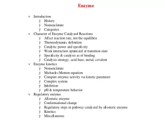
X Y C2 - Protein Functions: Enzymatic Enzyme:a protein*that catalyzes a chemical reaction, where a catalyst is defined as a substance that accelerates a chemical reaction without itself undergoing change. * some RNA molecules can also be considered enzymes A A + B B C + D • Specificity • Accelerated reaction rates • Control (regulation) • Enzymes can only affect the rate (kinetics) of a reaction, they can not make a reaction more energetically favorable. • Enzymes can be saturated by substrate.
v = Vs (KM + s) Basics of Enzyme Kinetics Michaelis-Menton Kinetics - for a simple enzyme reaction, the interaction of enzyme and substrate is considered an equilibrium and the overall reaction as follows: E + S ES E + P k+1 k-1 k+2 v = velocity, reaction rate KM = Michaelis constant KM = k2 + k-1 k1
C3 - Protein Functions: Signal Transduction Signal Transduction - in general the relaying of a signal from one physical form to another - in biological terms, the process by which a cell responds to signals (can be intracellular, extracellular). Signal Transduction Input Output • Examples of ‘signals’ (inputs): • chemicals • light • temperature • electrical (ion gradients) • other cells (cell-cell contact) • mechanical sensing
Generalized Model of Response to Extracellular Signal Ligand Activated Receptor Receptor “Action” • Ligand can activate or inactivate receptor • Output (action) dependent on system and sometime cell type • In metazoans (multi-cellular eukaryotes), there are about 16 intercellular classes of signaling systems
Example 1: Transmembrane Tyrosine Kinase Receptors Ligand P~ ~P Activated Receptor Receptor “Action” • Ligand binding results in receptor dimerization • The cytoplasmic (intracellular) domains are tyrosinekinases which phosphorylate each other on Tyr residue side chains. • This sets off a series of intracellular events
Example 2 : Steroid Receptors Ligand Activated Receptor Receptor nucleus • The steroid binds to it’s receptor in the cytoplasm. • The steroid-receptor complex but not the free receptor can move into the nucleus . • The steroid-receptor complex binds to specific binding site(s) on the DNA to regulate gene expression.
Example 3. Heterotrimeric G-Proteins Ligand Activated Receptor GTP GDP GDP Receptor GTP GTP (a b g complex) • Ligand binding causes activation of the a subunit which promotes exchange of GDP for GTP • In the GTP form, the a subunit and the associated bg subunits dissociate from the complex. • Each subunit can go on to initiate a series of intracellular events.
D - Regulation of Protein Activity The concentration of a protein in the cell is a function of the rate of synthesis and the rate of degradation. Both these processes can be regulated. Synthesis Transcription Translation Degradation DNA RNA Protein Proteins are often regulated such that the ‘activity’ of a protein is not a constant function of its concentration. Protein Active Protein Inactive
Regulation of Enzyme Activity Negative Feedback (Product Inhibition) X A B X A B C D E F Mechanistically negative feedback can be by direct competition of the product with the substrate for the active site or it can be indirect through interaction wit the enzyme away from the active site.
Regulation of Enzyme Activity Positive Feedback (Product Inhibition) X A B Positive Feedforward X A B
+ + Cooperativity / Allosteric Regulation Hypothetical examples of binding of a ligand to a dimeric protein. The binding curve is very sensitive to the effects on one site on the other. Two independent sites
+ + + + Cooperativity / Allosteric Regulation Hypothetical examples of binding of a ligand to a dimeric protein. The binding curve is very sensitive to the effects on one site on the other. Two independent sites Positive cooperativity
+ + + + + + Cooperativity / Allosteric Regulation Hypothetical examples of binding of a ligand to a dimeric protein. The binding curve is very sensitive to the effects on one site on the other. Two independent sites Positive cooperativity Negative cooperativity