Download
1 / 49
570 likes | 1.86k Views
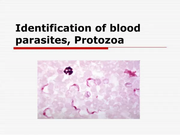
Diagnosis of Internal Parasites of Ruminants. Signs of Heavy Parasitism:

E N D
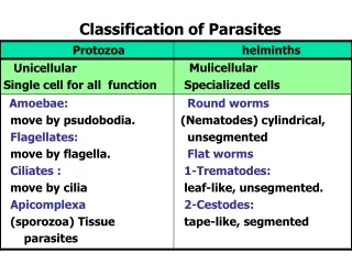
Signs of Heavy Parasitism: Infection with ruminant gastro-intestinal parasites may cause the following clinical signs to some degree, depending on the severity of infection: Diarrhea, dehydration, reduced weight gain and emaciation (1) bottle jaw (hypoproteinemia), rough hair root (2) and anemia (3). Liver and lung parasites may cause similar or additive effects involving liver and lung pathology respectively. Cattle and sheep have a striking similarity in the complement of parasites that affect them and usually only different species of identical genera are involved. In general, sheep are somewhat less resistant to the effects of parasitism than cattle and build heavier infection levels more easily. Only genera are discussed in this program.
Abomasal Parasites (Ostertagia, Haemonchus, Trichostrongylus axel)
Ostertagia egg Ostertagia has an egg that is morphologically typical of Trichostrongylid eggs. With only minor or statistical variations in size and shape, similar appearing eggs are also passed by Haemonchus, Trichostrongylus, CooperiaandOesophagostomwn. For reporting purposes, these eggs will be reported as trichostrongyle-type eggs by Diagnostic Services at I.SU. These are the most common type of eggs encountered in bovine fecal specimens and are reported as eggs per gram (EPG) after a quantitative sugar fecal flotation procedure
Ostertagia nodular lesions in abomasum. Ostertagia is probably the most important G-I parasite of cattle and sheep in Louisiana and causes small nodular lesions due to a .histotropic phase. of development in the abomasal mucosa.
Histotropic phase Each nodule contains a developing or an arrested. larvae which will eventually emerge to the lumen and the adult phase. In sufficient numbers these nodules and emergence of larvae are quite pathogenic, leading to disturbed mucosal secretory function, a rise in abomasal pH and loss of fluids and protein to the lumen. (fype II or Type I Ostertagiasis).
Adult Ostertagia compared to a paper clip Adult worms are small and are visible to the naked eye only with difficulty when recovered at necropsy.
Haemonchus contortus egg: Note similar appearance to Ostertagiaeggs. Haemonchus is a larger worm than Ostertagia (female 2.5cm; male 1cm: VS female 1.0cm ;male 0.5cm.).
Trichostrongylus eggs: Note typical trichostrongylid egg. Trichostrongylus often has a slightly”eccentric egg” shape as shown here. In sufficient numbers, this parasite may cause catarrhal inflammation and diarrhea (called the .bankrupt worm.). Trichostrongylus is a very small worm which is barely visible to the naked eye. (0.3- 0.5 cm).
Intestinal Parasites (Trichostrongylus, Cooperia, Nematodirus, Bunostomwn, Oesophagostomwn, Chabertia, Trichuris , Strongyloides , Moniezia
Cooperia ova: A typical “trichostrongylid” egg , note the longer shape with parallel sides as compared to other species. Intestinal Trichostrongylus spp. and Chabertia shed similar eggs.
Nematodirus in situ intestines: This long, thin worm (1-2 cm) is mainly found where cold climates prevail (it requires prolonged cold for development). Cooperia and Trichostrongylus are the most common intestinal nematodes in Southern states.
Nematodirus egg: Nematodirus has a very large unique egg which is not confusible with other trichostrongyles
Bunostomum in situ A hookworm that frequently infects cattle in Louisiana and warm climates {less frequently elsewhere). It is a voracious blood sucker and is found in the small intestine.
Bunostomum sp.: Bunostomum eggs are distinctive and are somewhat larger (80 x 50 u) and darker than trichostronylid eggs and should be reported as a separate item
Strongyloides papillosis egg: This species of cattle and sheep affects mainly young animals. It is called the “intestinal threadworm”(found in small intestine) and distinctive embryonated eggs are passed. Transmammary infection can occur .
Chabertia: This species (1-2cm) has a very large buccal capsule, skin penetration and inhabits the colon and is of relatively little importance.
Chabertia eggs: Typical “trichostrongylid “ egg
Oesophagostomum adults in situ / colon: Adults are large, stout worms (1-2cm) and can be a major pathogen in high numbers. The egg is a typical “trichostrongylid – type” .
Oesophagostomum lesions : The main damage of this species is by the larvae in the gut wall which produces small abcesses, mucosal sloughing.
Trichuris egg: Whipworms occur in the caecum of cattle and sheep and of little importance. Capillaria eggs from adults in small intestine (also of little importance) are also occasionally seen on fecal examinations
Eimeria sp. : Coccidia can cause bloody diarrhea in cattle or sheep particularly in feedlot or other close confinement situations, such as dairy calf barns. Although many species of Eimeria affect cattle and sheep, only a few are actually of any significance as pathogenic. In cattle: E. bovis, E. zurni are reported as “mixed coccidia including E. bovis, E. arlongi, etc.” Impression smears may be indicated on necropsy cases. Pathogenic species are identified by shape and size of oocyst.
Bovine rectum : Bloody diarrhea, lesions
Moniezia egg: This tapeworm of cattle and sheep is non-pathogenic. Note typical shape.
Mixed Parasite Infection -Flotation. Note relative sizes of “trichostrongyle-type “ eggs, Nematodirus , Moniezia and mixed Eimeria species. Moniezia Eimeria spp Strongyloides trichostrongyle-type Nematodirus
Comment of Quantitative egg counts. Quantitative egg counts will be used as a rough indicator of infection level. There are many variables (parasite species, fluid stool, .spring rise., .self-cure., immunity, age, etc.) that some say destroys any confidence put in quantitative counts in ruminants. With these limitations in mind, however, eggs per gram reporting can be a supporting factor in diagnosis parasitism. Several daily counts or a sample of at least 10 head from a herd tends to cancel variation. A mail-in fecal examination kit program is available to practitioners/county agents to monitor efficacy of parasite control programs (submit and mid winter example for nematodes; fall sample for flukes) herd history and anthelmintics used are considered in telephone advisories.
Necropsy Diagnosis At necropsy, parasites present can be identified to genera fairly easily by certain key traits. If a case is important enough to warrant the time necessary , a total worm count is the most reliable way to assess herd parasitism
Ostertagia -small ( < 1 cm).Abomasal worm with cervical papillae. Cervical papillae
Haemonchus -larger (1-3 cm) worms, barber pole appearance, cervical papillae present. Cervical papillae
Cooperia -alae at head, “railroad track” cuticular striations.
Trichostrongylus : “cervical notch”. Cervical notch
Nematodirus : male with very long, slender spicules. Other species with stout spicules. Females have “spine” at posterior end
Oesophagostomum -Note cervical alae, carona radiata; large robust size.
Fasciola egg: Large animal flukes eggs do not float and sedimentation procedures must be used.
Fasciola in situ/ bile ducts: Chronic wasting, anemia results from heavy infections.
Paramphistome. Rumen flukes; note conical morphology .
Paramphistome egg : Not distinguishable from Fasciola in, size or appearance.
Fascioloides magna : Common in Louisiana or swampy ranges shared with deer. Fluke host is normally deer; in cattle they wall off so no eggs escape to feces (dead end host). Characteristic very black pigment is often present.
Fascioloides magna adult.Note large robust shape with no -shoulders
Dicrocoelium : Small bile duct fluke which is endemic in New York. Intermediate host is black ant .
Dicrocoelium egg: Very small embryonated eggs are passed.
Thysanosoma in bile ducts of sheep: This “fringed tapeworm” occurs in high mountains (>6000 ft), in Western U.S. serving as a cause of condemned livers. “Fringe” on each segment of ThysanosomaCharacteristic eggs are passed
“Fringe” on each segment of Thysanosoma : Characteristic eggs are passed
Dictyocaulus first stage larva : Straight-tailed Ll stage is passed in stool. They will float (ZnSO4 spg 1.18 is the best flotation solution to use), but crenate if stand for long in solution. Baermannization can be done.
Muellaris / Protostrongylus : These lungworms occur in goats and sheep, producing blister1ike, granulomatous lesions beneath the serosal surface. Land snail is intermediate host
Muellerius larvae. Notch-tail larvae are produced and can be recovered by flotation ( ZnSO4 spg 1.18 works best) or Baermann apparatus.