Cell Membrane Structure and Function
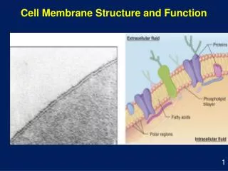
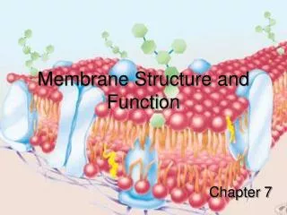
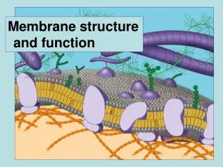
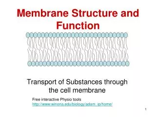
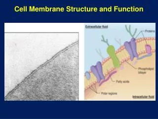
Cell Membrane Structure and Function. Membranes and Cell Transport. All cells are surrounded by a plasma membrane. Cell membranes are composed of a lipid bilayer with globular proteins embedded in the bilayer.

Cell Membrane Structure and Function
E N D
Presentation Transcript
Membranes and Cell Transport • All cells are surrounded by a plasma membrane. • Cell membranes are composed of a lipid bilayer with globular proteins embedded in the bilayer. • On the external surface, carbohydrate groups join with lipids to form glycolipids, and with proteins to form glycoproteins. These function as cell identity markers.
Fluid Mosaic Model Extracellular fluid Glycoprotein Glycolipid Carbohydrate Cholesterol Transmembrane proteins Peripheral protein Cytoplasm Filaments of cytoskeleton • In 1972, S. Singer and G. Nicolson proposed the Fluid Mosaic Model of membrane structure
Phospholipid Bilayer Polar hydro-philic heads Nonpolar hydro-phobic tails Polar hydro-philic heads • Mainly 2 layers of phospholipids; the non-polar tails point inward and the polar heads are on the surface. • Contains cholesterol in animal cells. • Is fluid, allowing proteins to move around within the bilayer.
Phospholipids Choline Hydrophilic head Phosphate Glycerol Hydrophobic tails Fatty acids Hydrophilic head Hydrophobic tails Space-filling model Structural formula Phospholipid symbol • In phospholipids, two of the –OH groups on glycerol are joined to fatty acids. The third –OH joins to a phosphate group which joins, in turn, to another polar group of atoms. • The phosphate and polar groups are hydrophilic (polar head) while the hydrocarbon chains of the 2 fatty acids are hydrophobic (nonpolar tails).
Membrane Components Cholesterol • Membrane carbohydrates • Interact with the surface molecules of other cells, facilitating cell-cell recognition • Cell-cell recognition is a cell’s ability to distinguish one type of neighboring cell from another • Steroid Cholesterol • Wedged between phospholipid molecules in the plasma membrane of animal cells. • At warm temperatures (such as 37°C), cholesterol restrains the movement of phospholipids and reduces fluidity. • At cool temperatures, it maintains fluidity by preventing tight packing. • Thus, cholesterol acts as a “temperature buffer” for the membrane, resisting changes in membrane fluidity as temperature changes.
Membrane Components Fibers of extracellular matrix (ECM) EXTRACELLULAR SIDE Glycoprotein N-terminus Carbohydrate Glycolipid Microfilaments of cytoskeleton C-terminus Peripheral protein Cholesterol Integral protein CYTOPLASMIC SIDE a Helix • Membrane Proteins • A membrane is a collage of different proteins embedded in the fluid matrix of the lipid bilayer • Peripheral proteins are appendages loosely bound to the surface of the membrane • Integral proteins penetrate the hydrophobic core of the lipid bilayer • Many are transmembrane proteins, completely spanning the membrane
Functions of Cell Membranes • Regulate the passage of substance into and out of cells and between cell organelles and cytosol • Detect chemical messengers arriving at the surface • Link adjacent cells together by membrane junctions • Anchor cells to the extracellular matrix
Functions of Plasma Membrane Proteins Outside Plasma membrane Inside Transporter Enzyme Cell surface receptor Cell surface identity marker Attachment to the cytoskeleton Cell adhesion
6 Major Functions Of Membrane Proteins ATP Enzymes Signal Receptor • Transport. (left) A protein that spans the membrane may provide a hydrophilic channel across the membrane that is selective for a particular solute. (right) Other transport proteins shuttle a substance from one side to the other by changing shape. Some of these proteins hydrolyze ATP as an energy source to actively pump substances across the membrane • Enzymatic activity. A protein built into the membrane may be an enzyme with its active site exposed to substances in the adjacent solution. In some cases, several enzymes in a membrane are organized as a team that carries out sequential steps of a metabolic pathway. • Signal transduction. A membrane protein may have a binding site with a specific shape that fits the shape of a chemical messenger, such as a hormone. The external messenger (signal) may cause a conformational change in the protein (receptor) that relays the message to the inside of the cell.
Glyco protein 6 Major Functions Of Membrane Proteins 4. Cell-cell recognition. Some glycoproteins serve as identification tags that are specifically recognized by other cells. 5. Intercellular joining. Membrane proteins of adjacent cells may hook together in various kinds of junctions, such as gap junctions or tight junctions Attachment to the cytoskeleton and extracellular matrix (ECM). Microfilaments or other elements of the cytoskeleton may be bonded to membrane proteins, a function that helps maintain cell shape and stabilizes the location of certain membrane proteins. Proteins that adhere to the ECM can coordinate extracellular and intracellular changes 6.
Cell Junctions • Long-lasting or permanent connections between adjacent cells, 3 types of cell junctions: • Tight junction • Anchoring junction • Communicating junction
Tight Junctions • Connect cells into sheets. Because these junctions form a tight seal between cells, in order to cross the sheet, substances must pass through the cells, they cannot pass between the cells. Tight junction
Anchoring Junctions Plasma membranes Intracellular attachment proteins Cell 1 Cell 2 Cytoskeletal filament Inter- cellular space Transmembrane linking proteins Extracellular matrix • Attach the cytoskeleton of a cell to the matrix surrounding the cell, or to the cytoskeleton of an adjacent cell.
Communicating (Gap) Junctions Two adjacent connexons form a gap junction Connexon Adjacent plasma membranes Intercellular space • Link the cytoplasms of 2 cells together, permitting the controlled passage of small molecules or ions between them.
Membrane Carbohydrates • Membrane carbohydrates interact with the surface molecules of other cells, facilitating cell-cell recognition • Cell-cell recognition is a cell’s ability to distinguish one type of neighboring cell from another
Membrane Transport • The plasma membrane is the boundary that separates the living cell from its nonliving surroundings • In order to survive, A cell must exchange materials with its surroundings, a process controlled by the plasma membrane • Materials must enter and leave the cell through the plasma membrane. • Membrane structure results in selective permeability, it allows some substances to cross it more easily than others
Passive Transport • Passive transport is diffusion of a substance across a membrane with no energy investment • 4 types • Simple diffusion • Dialysis • Osmosis • Facilitated diffusion
Diffusion • The net movement of a substance from an area of higher concentration to an area of lower concentration; down a concentration gradient • Caused by the constant random motion of all atoms and molecules • Movement of individual atoms & molecules is random, but each substance moves down its own concentration gradient.
Solutions and Transport • Solution – homogeneous mixture of two or more components • Solvent – dissolving medium • Solutes – components in smaller quantities within a solution • Intracellular fluid – nucleoplasm and cytosol • Extracellular fluid • Interstitial fluid – fluid on the exterior of the cell within tissues • Plasma – fluid component of blood
Diffusion Random movement leads to net movement down a concentration gradient Lump of sugar Water No net movement at equilibrium
Diffusion Across a Membrane Equilibrium Net diffusion Net diffusion • The membrane has pores large enough for the molecules to pass through. • Random movement of the molecules will cause some to pass through the pores; this will happen more often on the side with more molecules. The dye diffuses from where it is more concentrated to where it is less concentrated • This leads to a dynamic equilibrium: The solute molecules continue to cross the membrane, but at equal rates in both directions.
Diffusion Across a Membrane • Two different solutes are separated by a membrane that is permeable to both • Each solute diffuses down its own concentration gradient. • There will be a net diffusion of the purple molecules toward the left, even though the total solute concentration was initially greater on the left side Equilibrium Net diffusion Net diffusion Net diffusion Equilibrium Net diffusion
The Permeability of the Lipid Bilayer • Permeability Factors • Lipid solubility • Size • Charge • Presence of channels and transporters • Hydrophobic molecules are lipid soluble and can pass through the membrane rapidly • Polar molecules do not cross the membrane rapidly • Transport proteins allow passage of hydrophilic substances across the membrane
Passive Transport Processes • 3 special types of diffusion that involve movement of materials across a semipermeable membrane • Dialysis/selective diffusion of solutes • Lipid-soluble materials • Small molecules that can pass through membrane pores unassisted • Facilitated diffusion - substances require a protein carrier for passive transport • Osmosis – simple diffusion of water
Osmosis • Diffusion of the solvent across a semipermeable membrane. • In living systems the solvent is always water, so biologists generally define osmosis as the diffusion of water across a semipermeable membrane:
Lower concentration of solute (sugar) Higher concentration of sugar Same concentration of sugar Selectively permeable mem- brane: sugar mole- cules cannot pass through pores, but water molecules can Water molecules cluster around sugar molecules More free water molecules (higher concentration) Fewer free water molecules (lower concentration) Osmosis Water moves from an area of higher free water concentration to an area of lower free water concentration
Osmotic Pressure • Osmotic pressure of a solution is the pressure needed to keep it in equilibrium with pure H20. • The higher the [solutes] in a solution, the higher its osmotic pressure. • Tonicity is the ability of a solution to cause a cell to gain or lose water – based on the concentration of solutes
Fofi’s Definition of Osmosis Osmosis is the diffusion of water across a semipermeable membrane from a hypotonic solution to a hypertonic solution
Tonicity Hypotonic solution Hypertonic solution Isotonic solution H2O H2O H2O H2O Normal Shriveled Lysed • If 2 solutions have equal [solutes], they are called isotonic • If one has a higher [solute], and lower [solvent], is hypertonic • The one with a lower [solute], and higher [solvent], is hypotonic
Facilitated Diffusion • Diffusion of solutes through a semipermeable membrane with the help of special transport proteins i.e. large polar molecules and ions that cannot pass through phospholipid bilayer. • Two types of transport proteins can help ions and large polar molecules diffuse through cell membranes: • Channel proteins – provide a narrow channel for the substance to pass through. • Carrier proteins – physically bind to the substance on one side of membrane and release it on the other.
Facilitated Diffusion • Specific – each channel or carrier transports certain ions or molecules only • Passive – direction of net movement is always down the concentration gradient • Saturates – once all transport proteins are in use, rate of diffusion cannot be increased further
Active Transport • Uses energy (from ATP) to move a substance against its natural tendency e.g. up a concentration gradient. • Requires the use of carrier proteins (transport proteins that physically bind to the substance being transported). • 2 types: • Membrane pump (protein-mediated active transport) • Coupled transport (cotransport).
Membrane Pump • A carrier protein uses energy from ATP to move a substance across a membrane, up its concentration gradient:
The Sodium-potassium Pump • One type of active transport system [Na+] high [K+] low Na+ Na+ 1. Cytoplasmic Na+ binds to the sodium-potassium pump. 2. Na+ binding stimulates phosphorylation by ATP. Na+ Na+ EXTRACELLULAR FLUID Na+ ATP [Na+] low [K+] high P Na+ ADP CYTOPLASM Na+ Na+ 6. K+ is released and Na+ sites are receptive again; the cycle repeats. 3. Phosphorylation causes the protein to change its conformation, expelling Na+ to the outside. Na+ K+ P K+ 5. Loss of the phosphate restores the protein’s original conformation. K+ 4. Extracellular K+ binds to the protein, triggering release of the Phosphate group. K+ K+ K+ P i P P i
Coupled transport 2 stages: • Carrier protein uses ATP to move a substance across the membrane against its concentration gradient. Storing energy. • Coupled transport protein allows the substance to move down its concentration gradient using the stored energy to move a second substance up its concentration gradient:
Bulk Transport • Allows small particles, or groups of molecules to enter or leave a cell without actually passing through the membrane. • 2 mechanisms of bulk transport: endocytosis and exocytosis.
Endocytosis • The plasma membrane envelops small particles or fluid, then seals on itself to form a vesicle or vacuole which enters the cell: • Phagocytosis • Pinocytosis
Phagocytosis The substance engulfed is a solid particle
Pinocytosis Thesubstance engulfed is a liquid
Exocytosis • The reverse of endocytosis • During this process, the membrane of a vesicle fuses with the plasma membrane and its contents are released outside the cell:
Cells Communication • Direct contact • Paracrine signaling • Endocrine signaling • Synaptic signaling
Direct Contact Adjacent connexons form a gap junction • Cells touch each other and signal molecules travel through special connections called communicating junctions • Communicating Junctions link the cytoplasms of 2 cells together, permitting the controlled passage of small molecules or ions between them.
Local and long-distance cell communication in animals Local signaling Long-distance signaling Blood vessel Endocrine cell Target cell Electrical signal along nerve cell triggers release of neurotransmitter Neurotransmitter diffuses acrosssynapse Secretory vesicle Secreting cell Hormone travels in bloodstream to target cells Local regulator diffuses through extracellular fluid Target cell Target cell is stimulated (b) Synaptic signaling. A nerve cell releases neurotransmitter molecules into a synapse, stimulating the target cell. (a) Paracrine signaling. A secreting cell acts on nearby target cells by discharging molecules of a local regulator (a growth factor, for example) into the extracellular fluid. (c) Hormonal signaling. Specialized endocrine cells secrete hormones into body fluids, often the blood. Hormones may reach virtually all body cells.
Cell Signaling EXTRACELLULAR FLUID CYTOPLASM Plasma membrane 3 1 2 Reception Transduction Response Receptor Activation of cellular response Relay molecules in a signal transduction pathway Signal molecule • The cells of a organism communicate with each other by releasing signal molecules that bind to receptor proteins located either on or inside of target cells. • Three stages of cell signaling: • Reception - each target cell has receptors that detect a specific signal molecule and binds to it • Transduction – binding of the signal molecule changes the receptor protein in some way that initiates transduction or conversion of the signal to a form that can bring about a specific cellular response • Response – transduced signal triggers a specific cellular response, any cell activity
Reception • A signal molecule binds to a receptor protein, causing it to change shape • The binding between signal molecule (ligand) and receptor is highly specific • A conformational change in a receptor • Often the initial transduction of the signal
Cell Receptors • Cell surface receptors. - Signal molecules that cannot pass through the plasma membrane bind to receptors located on the surface of the membrane • Chemical or ligand-gated ion channels • Enzymatic receptors • G-protein-linked receptors • Intracellular receptors • Some signal molecules that are small or hydrophobic can pass through the plasma membrane and bind to receptors located inside the cell • Intracellular receptors are cytoplasmic or nuclear proteins
Intracellular Receptors • Gene Regulators • Signal molecule joins to the receptor, the receptor changes shape and a DNA binding site is exposed. • The DNA binding site joins to a specific segment of DNA and activates (or suppresses) a particular gene • Enzyme Receptor • These receptors function as enzymes – proteins that catalyze (speed up) specific chemical reactions. • When a signal molecule joins to the receptor, the receptor’s catalytic domain is activated (or deactivated).
Steroid hormone interacting with an intracellular receptor Hormone (testosterone) EXTRACELLULAR FLUID The steroid hormone testosterone passes through the plasma membrane. 4 3 2 5 1 Plasma membrane Testosterone binds to a receptor protein in the cytoplasm, activating it. Receptor protein Hormone- receptor complex The hormone- receptor complex enters the nucleus and binds to specific genes. DNA The bound protein stimulates the transcription of the gene into mRNA. mRNA NUCLEUS New protein The mRNA is translated into a specific protein. CYTOPLASM