Download
1 / 49
500 likes | 956 Views
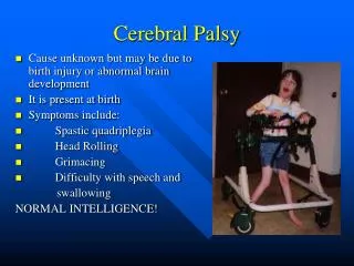
CEREBRAL PALSY. Presenter – Dr.Sudhanshu Kothadia. Defination. “cerebral Palsy Is A Disorder of posture & Movement Secondary To Static Lesion At The Developing Brain.”. The word cerebral refers to the brain's two halves, or hemispheres.

E N D
CEREBRALPALSY Presenter – Dr.SudhanshuKothadia
Defination “cerebral Palsy Is A Disorder of posture & Movement Secondary To Static Lesion At The Developing Brain.” DR.SUDHANSHU
The word cerebral refers to the brain's two halves, or hemispheres. • The word palsy refers to any disorder that impairs control of body movement.
CLASSIFICATION The widely accepted classification of cerebral palsy described by minear (1956) includes 7 major categories , 34 minor categories & numerous subcategories. Based On Motor Involvement: 1. Spastic C.P This is the most frequent type of cerebral palsy. There is increased resistance to passive movements. There is first initial resistance & after that there may be relaxation known as jack knife spasticity. It is present in pyramidal tract, usually in motor cortex. DR.SUDHANSHU
2.Involuntary movements • Involuntary Activity Is Accentuated By Emmotional Activity Occurs In The Lesion At Basal Ganglia. There are two types:- • Chorioathetoid • Dystonic • Chorioathetoid is a combination of choreiac & athetoid. • Choreiac movements are gross & fast arrhythmic with sudden beginning. • Athetoid are continuous , uniform & slow. • Dystoniacharacterised by intermittent twisting movements dystonic movements commonly determine bizzare posture. DR.SUDHANSHU
3. Rigidity This also occurs due to lesion in basal ganglia. It causes generalisedhypertonia secondary to continuous contraction of flexor extensor muscles. 4.Ataxia It is characterised by clumsy gaitwith broadening of the base due to lack of balance . Ataxia mainly related to cerebellar disorders & rarely occurs with cerebral palsy . 5.Hypotonia Rarely occurs with cerebral palsy. DR.SUDHANSHU
Classification based on Tomography 1. Monoplegia Rare condition in cerebral palsy Involves only one limb 2. Hemiplegia Involves one half side of body Commonly present with cerebral palsy 3. Paraplegia If the child has pure paraplegia then it will be considered as pure to medulla lesion DR.SUDHANSHU
4.Diplegia Upper & lower extremities are involved It is commonly related to prematurity 5. Triplegia There is involvement of three limbs Generally two legs & one arm DR.SUDHANSHU
Classification based on Severity 1. Mild:-fine movements alterations only 2. Moderate:-variable difficulty in elation to speech & gross movements 3. Severe:-inability to use hands, to walk & to communicate DR.SUDHANSHU
Etiology Prenatal Perinatal Postnatal Prenatal:- Congenital infections Toxoplasmosis, Rubella, Cytomegalovirus Abuse of drugs Tobbaco, Alcohol, Marijuana, Cocaine Cerebral malformations Obstetric complications Pre-eclampsia/Eclampsia, Abrutioplacentae, Placenta previa DR.SUDHANSHU
Perinatal Premaurity Low birth weight Comlpicated delivery (dystocia) Asphyxia, Cerebral trauma Infection Meningitis, Herpes Hyperbilirubinaemia Blood incompatibilitis, Other haemolytic disorders Hypoglycaemia DR.SUDHANSHU
Postnatal Infections Meningitis, Encephalitis Head trauma Cerebrovascular accidents Cynotic congenital heart Disease, Sickel cell anaemia, Vascular malformations Cerbral anoxia Near drowning, Aspiration asphyxia, Cardiac arrest, seizures malnutrition DR.SUDHANSHU
Clinical Presentation • Remember that motor developmental progression is from….Head to Toe
Diagnosis Diagnosis of C.P. is difficult to diagnose under the age of 12 months until deformity develops. What usually brings he attention is motor delay. In a child with hypotonia, hypo or arreflexia, apparent muscular atrophy, absence of involuntary movements & good cognitive development, the disorder must be due to lower motor neuron. DR.SUDHANSHU
Umn lesion may be located in cerebral cortex, internal capsule, cerebral peduncles, brain stem or spinal cord& are associated with pyramidal signs. Both hypertonia & hyperreflexia reflects Umn lesion. In the first months of life , sucking or swallowing difficulties & neck hyperextension are strongly suggestive of a cerebral lesion. Inspite of the fact that C.P. is non progressive, there will be changes in neurological picture due to CNS maturation. DR.SUDHANSHU
Prognostic Factors Considerable Work has been done to investigate prognostic factors. Paine et all noted that sitting independently by 2 yrs old was a good predictor of independent ambulation. He found that approximately half of children who can sit independently by 2 to 4 yrs can eventually walk and if child cant sit independently by 4 yrs it is unlikely that he or she will walk without support. Finally if a child has not learned to walk by 8 yrs and if he or she is not limited by severe contractures it is unlikely that she will ever walk. DR.SUDHANSHU
Bleck also gave prognostic facors on the basis of primitive reflexes. Poor prognostic signs for walking includes : 1) An imposable asymmetrical tonic neck reflex 2) Persistent Moro’s reflex 3) Strong Extensor Thrust on Vertical Suspension 4) Absence of Normal Parachute reaction beyond 11 months. DR.SUDHANSHU
MANAGEMENT REHABILITATION:- Patient training Communication Seating Walking & Mobility Bracing Main indications of bracing in cerebral palsy are:- Early post operative stages to induce early walking. Following G.A. manipulation & Myoneuaral blocking to post pone tendon lengthening. DR.SUDHANSHU
Treatment Non –operative Operative DR.SUDHANSHU
Non-operative • Non-operative modalitis includes medication , splinting & bracing, physical therapy. • Variety of medication are used, common are- • Diazepam – Acts centrally • Baclofen – Acts centrally • Dantrolene-Acts at the level of skeletal muscle. • As these drugs increases the neurotransmitter activity, there has common systemic side effects such as sedation, balance difficulties & cognitive dysfunction. DR.SUDHANSHU
Dantrolene Dantrolene acts at the level of skeletal muscle & decreases muscle calcium iron release. It has affinity for fast twitch muscle fibres & selecively decreases abnormal muscle stretch reflex & tone. It has side effect as it can cause weakness & hepatotoxicity on prolonged use. DR.SUDHANSHU
Baclofen It inhibits mono synaptic extensor activity & poly synaptic flexor activity and it has been shown to decrease substance P level. As these have poor BBB penetration capacity, high doses are required. Intrathecal injection requires 1/30th dose of baclofen to achieve optimum effect. Side effects of baclofen includes :- Catheter or pump infection Respiratory infection Drug reaction Oversedation DR.SUDHANSHU
Botulinum Toxin It is a potent neurotoxin of which there are seven serotypes, produced by clostridium botulinum. Botulinum toxin –A (BTX-A) has been used to weaken the muscle spasticity. BTX-A injected directly into muscle act at motor end plate inhibiting release of acetylcholine, therefore inhibiting muscle contraction. Effects begins 24 hrs after injection & lasts for 2-6 months. The maximal safe dose of BTX-A is 36-50 U/kg. High dose can cause respiratory depression & death. BTX-A is more effective when used with other modalities such as physical therapy & serial casting. DR.SUDHANSHU
Contraindications of Botulinum Toxin:- Known resistant or antibodies. Fixed deformities or contractures. Concurrent use of Aminoglycosides. Failure of previous response. Certain neurological condition such as myesthenia gravis. DR.SUDHANSHU
Physical Therapy It is also an essential component of treatment. It is typically used as primary treatment in conjunction with BTX-A injection, casting, splinting, bracing & surgery. DR.SUDHANSHU
Bracing Used mainly to prevent or slow the progression of deformity. Most common brace used in C.P. is ankle-foot arthrosis, hip adduction braces, hand & wrist splints, spinal braces & jackects. DR.SUDHANSHU
Operative Treatment Indicated when contracture & deformity decrease the function, cause pain or interfere with the activities of daily living. Operative treatment can be divided further, including procedures to:- Correct static or dynamic deformities. Balances muscle power across the joint. Releases spasticity (neurerectomy). Stabilize uncontrollable joints. Flexible static & dynamic deformities are corrected by muscle tendinious lengthening. Capsulotomy & osteotomies are reserved for more severe & rigid deformities. DR.SUDHANSHU
Regional Deformities & their management DR.SUDHANSHU
Hip Deformities around hip occurs from painless subluxation to painful dislocation. In most of the patients, hip is normal at birth & radiological changes clinically evident between 2-4 yrs. The cause of this progressive deformity is multifactorial & includes muscle imbalance, retained primitive reflexes, abnormal positioning & pelvic obliquity. DR.SUDHANSHU
Some deformities are :- Acetabular dysplasia, increased neck shaft angle, increased anteversion. Increased neck shaft angle is common in ambulators. Neck shaft angle increases in age in children with C.P. rather to decrease as normally. Anteversion which commonly decreases with age does not change in C.P. children. DR.SUDHANSHU
Flexion Deformities of Hip These are common in C.P. Excessive flexion at hip joint produces crouch gait. Excessive flexion causes shifting of centre of gravity anteriorly compensated by lumbar lordosis , knee flexion & ankle dorsiflexion causing crouch gait. A crouched gait in a child who has cerebral palsy may be caused by spasticity of the iliopsoas muscle with resultant hip-flexion deformity, by spasticity of the ham string muscles, by calcaneus deformity of the foot and ankle, or by a combination of these problems. Hip flexion deformities from 15-30 degrees usually treated by Psos lengthening through an intramuscular recession over pelvic brim. Contracture >30 requires extensive release of rectus femoris, sartorius, TFL & ant. Fibres of gluteus minimus & medius. DR.SUDHANSHU
Adduction deformities Most common deformity in C.P. in hip. For mild deformity adductor tenotomy is sufficient. For more severe deformity release of gracillis & ant. Head of adductor bravis required. Adductor tenotomy is usually bilateral & plaster is given to prevent recurrence. DR.SUDHANSHU
It is difficult to diffentiate between Adduction & pseudo adduction i.e. foot-internal rotation deformity. In pseudoflexion children sits with ‘W’ position i.e. -Hips are flexed at 90 degrees & internally rotated. -Knees are flexed & internally rotated. -Feets are externally rotated DR.SUDHANSHU
Subluxation & Dislocation Of Hip Hip dislocation occurs on continued mild subluxation. Maximum percentage of hip dislocation is in quadriplegic patients. Hip with flexion contractures of >20 degrees & abduction at > 30 degrees are of increasing risk of progressive subluxation. Varusderotationosteotomy used to correct hip dislocation. DR.SUDHANSHU
Knee Hip & Knee relationship:- • Rarely deformities occurs isolately in knee. • Generally hip & knee deformities occurs in conjunction because of the muscles that crosses the joints. • There includes:- • Rectus femorisanteriorly • Gracillis medially • Semi membranous, semitendinousposteriorly. DR.SUDHANSHU
A similar relationship exist between knee & ankle due to gastrocnemius. Develops Jump gait causing flexion at hip , knee and plantar flexion at foot. Corrected by hamstring tendon release & lengthening. Care should be taken to avoid overlengthening as it produces hyperextension gait. DR.SUDHANSHU
Foot EquinusDeformity:- Most common in C.P. Occurs due to spasticity of gastrocnemius, soleus which often worsens during periods at rapid growth because of overgrowth of tibia relative to gastrocnemius. Corrected by T.A. lengthening. DR.SUDHANSHU
Varus or valgus • Occurs with equinus. • Direction of deformity depends upon type & severity of C.P. • In hemiplegia,deformity is either equines or equivarus. • In diplegia or quadriplegia, valgus or equinovalgus. • Varus deformities are more functionally disability, difficult to manage Conservatively and easy to manage operatively. • Lengethening at posterior tibial tendon done to correct varus deformity. • Calcaneusosteotomy is used to correct to equinovalgus. DR.SUDHANSHU
Calcaneus Deformity Pure calcaneus deformity is rare and it is associated with calcaneovalgus. Caused due to overlengthening of achillis tendon. Corrected by cresentricosteotomy of calcaneus DR.SUDHANSHU
Another Rare Deformities Forefoot adduction Cavus Hallusvalgus Claw toes DR.SUDHANSHU
Spinal Deformities 1) Kyphosis 2) Lordosis 3) Scoliosis DR.SUDHANSHU
Kyphosis The most common type of kyphosis associated with cerebral palsy is seen in children who have quadni paretic involvement and substantial weakness of the spinal extensor muscles. Such patients have a long. Al most total c-shaped kyphosis that is characteristic of paralytic conditions. The apex is in the middle or lower thoracic area. The deformity almost always is flexible and corrects completely when the patient is placed in prone position. DR.SUDHANSHU
Treatment consists of modification of the wheelchair with straps that restrain the chest or with a harness to maintain an upright sitting posture. A total-contact type of thoracolumbosacral trunk-support orthosis usually is successful for patients who have severe problems operative stabilization of the spine with arthrodesis and internal fixation is necessary only for patients who have the most severe involvement. DR.SUDHANSHU
Kyphosis with the apex in the lumbar spine may occur when spastic hamstrings proximally pull the pelvis and lower lumbar spine into flexion. This deformity, which may interfere with stable sitting, usually can be corrected with a proximal release of the hamstrings'. DR.SUDHANSHU
Lordosis In patients who have cerebral palsy, hyperlordosis most often occurs in the lumbar spine as a consequence of substantial hip-flexion contractures and usually responds to lengthening of the hip flexors by either stretching or operative elongation. Thoracic or thoracolumbarhyperbordosis is common in patients who have scoliosis. If the scoliosis is treated operatively, it is important to evaluate the lardosis and to maintain appropriate balance in the sagital plane. DR.SUDHANSHU
Scoliosis Patients who have spasticity and quadriplegia are more likely to have scoliosis. Patients who have scoliosis in association with cerebral palsy can be managed with observation and documentation, an orthosis, or spinal arthrodesis with instrumentation. Observation alone is appropriate. An orthosis usually is ineffective for controlling or stopping the progression of scoliosis in patients who have cerebral palsy. DR.SUDHANSHU
Spinal arthrodesis with internal fixation is the definitive treatment of progressive scobiosis in patients who have cerebral palsy. Operative treatment is indicated if the scoliosis exceeds 40 to 50 degrees and if the spine will not be severely shortened by arrest of its further growth (usually after the patient has reached the age of nine or ten Yrs. DR.SUDHANSHU
THANK YOU DR.SUDHANSHU