Download

1 / 52
540 likes | 1.13k Views
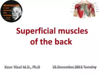
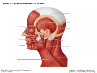
Superficial muscles of the back. Kaan Yücel M.D., Ph.D. 29.December.2011 Thursday. MUSCLES OF THE BACK Extrinsic back muscles Superficial group consists of muscles related to and involved in movements of the upper limb.

E N D
Superficial muscles of the back Kaan Yücel M.D., Ph.D 29.December.2011 Thursday
MUSCLES OF THE BACK • Extrinsic back muscles • Superficial group consists of muscles related to and involved in movements of the upper limb. • Intermediate group consists of muscles attached to the ribs and may serve as a respiratory function. • Intrinsic (deep) back muscles • Act on the vertebral column, • producing its movements and • maintaining posture.
SUPERFICIAL GROUP OF BACK MUSCLES • Immediatelydeep to the skin and superficial fascia. • Connectedwith the shoulder girdle. • Attachthe superior part of the appendicular skeleton (clavicle, scapula, and humerus) to the axial skeleton (skull, ribs, and vertebral column). • Produceand control limb movements. • Trapezius • Latissimusdorsi • Rhomboid major • Rhomboid minor • Levator scapulae
Rhomboid major, rhomboid minor, and levator scapulae are located deep to trapezius in the superior part of the back. Although located in the back region, for the most part these muscles receive their nerve supply from the anterior rami of cervical nerves and act on the upper limb. The trapezius receives its motor fibers from a cranial nerve, the spinal accessory nerve (CN XI).
Flat & triangular, muscle's origin: base of triangle situated along vertebral column muscle's insertion: apex pointing toward tip of shoulder Covers posterior aspect of neck & superior half of trunk.
It was given its name because the muscles of the two sides form a trapezium (G. irregular four-sided figure). The muscles on both sides together form a trapezoid.
The trapezius attaches the pectoral girdle to the cranium and vertebral column and assists in suspending the upper limb.
The fibers of the trapezius are divided into 3 parts, • different actions at the physiological scapulothoracic joint between the scapula and the thoracic wall: • Descending (superior) fibers elevate the scapula • (e.g., when squaring the shoulders). • Middle fibers • retract the scapula • (i.e., pull it posteriorly). • Ascending (inferior) fibers depress the scapula and • lower the shoulder.
The superior fibers of trapezius, from the skull and upper portion of the vertebral column, descend to attach to the lateral third of the clavicle and to the acromion of the scapula.
Superior and inferior fibers work together to rotate the lateral aspect of the scapula upward, which needs to occur when raising the upper limb above the head. Descending and ascending trapezius fibers act together in rotating the scapula on the thoracic wall in different directions, twisting it like a wing nut.
The trapezius also braces the shoulders by pulling the scapulae posteriorly and superiorly, fixing them in position on the thoracic wall with tonic contraction; consequently, weakness of this muscle causes drooping of the shoulders.
Motor innervation of trapezius accessory nerve [XI] descends from the neck onto the deep surface of the muscle. Proprioceptive fibers from trapezius pass in the branches of the cervical plexus and enter the spinal cord at spinal cord levels C3 &C4.
Latissimus dorsi(L. widest of back) • Large, flat triangular muscle • Begins in the lower portion of the back • Tapers as it ascends to a narrow tendon that attaches to the humerus anteriorly.
Posterior axillary fold formed by the tendon of latissimus dorsi as it passes around the lower border of the teres major muscle. Easily palpated between the finger and thumb.
This large, fan-shaped muscle passes from the trunk to the humerus and acts directly on the glenohumeral joint and indirectly on the pectoral girdle (scapulothoracic joint).
The latissimus dorsi extends, retracts, and rotates the humerus medially (e.g., when folding the arms behind the back or scratching the skin over the opposite scapula). • As a result, movements associated with this muscle include • Extension • Adduction • Medial rotation of the upper limb • Latissimus dorsi can also depress the shoulder, preventing its upward movement.
In combination with the pectoralis major, the latissimus dorsi is a powerful adductor of the humerus and plays a major role in downward rotation of the scapula in association with this movement.
It is also useful in restoring the upper limb from abduction superior to the shoulder; hence the latissimus dorsi is important in climbing.
In conjunction with the pectoralis major, the latissimus dorsi raises the trunk to the arm, which occurs when performing chin-ups or climbing a tree, for example. These movements are also used when chopping wood, paddling a canoe, and swimming (particularly during the crawl stroke).
The superior third of the strap-like levator scapulae lies deep to the sternocleidomastoid; the inferior third is deep to the trapezius.
From the transverse processes of the upper cervical vertebrae, the fibers of the levator of the scapula pass inferiorly to the superomedial border of the scapula.
True to its name, the levator scapulae acts with the descending part of the trapezius to elevate the scapula, or fix it (resists forces that would depress it, as when carrying a load).
With rhomboids & pectoralis minor, rotates the scapula, depressing the glenoid cavity (rotating the lateral aspect of scapula inferiorly).
Acting bilaterally (also with the trapezius), the levators extend the neck. • Acting unilaterally, may contribute to lateral flexion of the neck (toward the side of the active muscle).
RHOMBOID MINOR & RHOMBOID MAJOR
The rhomboids (major and minor), which are not always clearly separated from each other, have a rhomboid appearance—that is, they form an oblique equilateral parallelogram. Lie deep to the trapezius, inferior to levator scapulae and form broad parallel bands that pass inferolaterally from the vertebrae to the medial border of the scapulae.
Rhomboid minor • superior to rhomboid major, • small, cylindrical muscle • Arises from ligamentum nuchae & spinous processes of vertebrae CVII and TI • Attaches to medial scapular border opposite root of spine of scapula.
The larger Rhomboid major Origin: Spinous processes of upper thoracic vertebrae Attaches: Medial scapular border inferior to rhomboid minor
Retract & rotate scapula Assist serratus anterior in holding the scapula against the thoracic wall and fixing the scapula during movements of the upper limb. Used when forcibly lowering the raised upper limbs (e.g., when driving a stake with a sledge hammer).
INTERMEDIATE GROUP OF BACK MUSCLES
2 thin muscular sheets in the superior and inferior regions of the back, immediately deep to the muscles in the superficial group. • Related to the movements of the thoracic cage, as the superficial muscles are related to the movements of the shoulder (girdle). • The intermediate extrinsic back muscles (serratus posterior) are thin muscles, commonly designated as superficial respiratory muscles, but are more likely proprioceptive rather than motor in function.
Described with muscles of the thoracic wall: Serratus posterior superior lies deep to the rhomboids Serratus posterior inferiorlies deep to the latissimus dorsi
Both serratus posterior muscles are attached to the vertebral column and associated structures medially Either descend (fibers of serratus posterior superior) or Ascend (fibers of serratus posterior inferior) to attach to the ribs. These two muscles therefore elevate and depress the ribs.