Download
1 / 1
10 likes | 107 Views
Measuring the galvanotaxic response of Amoeba proteus to a changing DC electrical field J. Bryce Sealander, Joseph Merriman, James K. Brown, Francis X. Hart PhD, John R. Palisano PhD Departments of Biology and Physics, The University of the South, 735 University Avenue, Sewanee, Tennessee 37383.

E N D
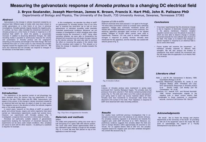
Measuring the galvanotaxic response of Amoeba proteus to a changing DC electrical field J. Bryce Sealander, Joseph Merriman, James K. Brown, Francis X. Hart PhD, John R. Palisano PhDDepartments of Biology and Physics, The University of the South, 735 University Avenue, Sewanee, Tennessee 37383 Abstract Galvanotaxis is the change in cellular movement created by an electrical field. Different types of motile cells have been shown to exhibit galvanotaxis on a time frame of an hour in previous investigations. Amoeba typically migrate towards the negative pole. In contrast, we have video recorded the directional movement of Amoeba proteus on a time frame of minutesin response to a DC electrical field (dcEF) for which the polarity is occasionally reversed. In addition to observing directional changes, the video recording makes it possible to continuously measure the velocity and acceleration of Amoeba proteus and the cytoplasmic streaming as the polarity and strength of the field change. We have confirmed that even on a time scale of a few minutes the amoeba migrates toward the negative pole in a field of about 300 V/m. We have also observed that the amoeba can respond to changes in the direction of the field within a minute. In this investigation, we describe the effect of dcEF on galvanotaxis on Amoeba proteus. One of our aims was to examine the short-term (up to half an hour) galvanotaxis of individual amoebae under precisely controlled experimental conditions. Moreover, we are unaware of investigations in which amoebae were video recorded during the movement in dcEF. Using video recording software, we were able to characterize the directed cell movement and cell shape changes induced by the electric stimulus. We were also able to examine the effect on reversing the polarity of the electric field that the amoebae were subject to. This allowed us to follow the change in migration of amoeba towards the negative pole. Exposure of Cells to dcEFs Platinum electrode wires were placed 3.1 cm apart in the trough. The wires were connected in series to a resistor and a switching apparatus. Voltage was continually monitored across the resistor with a Digital Multimeter to insure current continuity. The switching apparatus permitted rapid reversal of the applied polarity. Voltages of 5V-20V, direct current, were used in experiments. However, 10V was used to monitor migrational changes in response to polarity reversal.Amoeba were introduced into the apparatus, and the chamber was sealed with silicone grease (Fig. 3). • Conclusions • It has been hypothesized that this directional change is due to migration of electrically charged receptors in the plasma membrane. However, receptor migration cannot occur over the period of seconds in which we were able to observe change in amoeba movement towards the new cathode upon reversal of the field. This leads us to believe that changes in cellular migration in response to an electrical field is due to some other factor. • Future studies will examine the movement of individual amoeba exposed to different field strengths. We will also analyze the kinetics of cytoplasmic flow which controls the movement and the cell shape after the application of an electric field or its reversal. Literature cited Adler, J. and W. Shi.Galvanotaxis in Bactera.1988.Quantitative Biology 3: 23-25. Korohoda, Wlodzimierz, Mycielska, M., Janda, E. and Zbigniew Madeja. 2000. Immediate and Long- term Galvanotactic Responses of Amoeba proteus to dc Electric Fields. Cell Motility and the Cytoskeleton 45: 10-26. Nishimura, K.Y., Isseroff, R.R., and Richard Nuccitelli. 1996. Human keratinocytes migrate to the negative pole in direct current electric field comparable to those measured in mammalian wounds. Journal of Cell Science 109: 199-207. Fig. 4 Amoeba Culture Fig. 2 Diagram of slide preparation Cells Cultures of Amoeba proteus were maintained in spring water purchased from Carolina Biological Supply. The amoeba were fed protozoan that were cultured in Petri dishes containing boiled wheat seeds (Fig. 4). The cells were taken out of the culture and placed in pure spring water to wash any excess materials debris off them. Amoeba were then transferred to microscope slide where they attached to the surface of the slide. Their migrations in response to dcEF were observed with video recording software. Fig. 1 Amoeba proteus Introduction The importance of the electrical control of cell physiology has been stressed since the famous frog leg experiments of Luigi Galvani in the late 1700s (Adler and Shi 1988). Galvanotaxis, one aspect of this control, is the change in cellular movement created by an electrical field and has been an object of study for many years. This migration occurs in the direction of either the cathode (negative pole) or the anode (positive pole). In recent years, research on the effects of dcEF on growth of plants and animal tissues, organs and entire organisms have been carried out. Galvanotaxis experiments usually last several hours. However, our experiments with Amoeba proteus (Fig. 1) investigated a response that occurred on a time scale of minutes. Amoebae are particularly suitable subjects of galvanotactic experiments studying the mechanism of movement because the experiments can be done under simple conditions. Also, amoebae are easier to work with for other reasons – no plating on a substrate is required, for example. Results Our studies have confirmed previous investigations that in an isotropic environment and in the absence of any electric stimulus, the amoebae randomly migrated in all directions but towards the negative pole under dcEF (Korohod et al. 1997). Moreover, our preliminary evidence suggests that amoeba detect the field and start to change direction within approximately 45 seconds to one minute. For an applied electric field of 300 V/m, the amoeba migrated toward the negative pole and often exhibited elongation into a leech-like structure (Fig. 5). Fig. 3 Top View of Apparatus for Amoeba Acknowledgments We would like to thank the biology and physics departments of the University of the South for providing us with all of the equipment used in the study. We also want to acknowledge the support of a Faculty Development Fund Grant to JRP. Materials and methods Slides The slides were prepared by cutting one cover slip in half and gluing it to a glass slide with silicone sealant. The cover slips were placed approximately three to four millimeters apart creating a trough for the amoeba (Fig. 2). A cover slip was then placed on top of the apparatus to seal the trough.