
Managing Post-Radiotherapy Neck in Head and Neck Cancer: Physical Exam vs. Computed Tomography
A study comparing physical exams and CT scans in managing post-radiotherapy neck dissections for head and neck cancer patients. Results showed CT imaging was more effective in identifying residual disease.

Managing Post-Radiotherapy Neck in Head and Neck Cancer: Physical Exam vs. Computed Tomography
E N D
Presentation Transcript
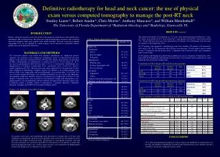
Definitive radiotherapy for head and neck cancer: the use of physical exam versus computed tomography to manage the post-RT neckStanley Liauw*, Robert Amdur*, Chris Morris*, Anthony Mancuso^, and William Mendenhall*The University of Florida Department of *Radiation Oncology and ^Radiology, Gainvesille FL RESULTS (updated) INTRODUCTION The median followup for all 550 patients was 3.3 years, while median potential followup was 7 years. At 5 years, neck control was 90%, local control 84%, cause-specific survival 69%, and overall survival 52%. 396 post-RT neck dissections were performed in 341 patients. Post-radiotherapy neck dissection was negative in 256 heminecks (67%). Of 537 patients (684 heminecks) with physical exam data available, 259 patients (435 heminecks, 64%) had a cCR. Of 142 heminecks with cCR that were dissected, 33 were found to have residual disease; 17 heminecks had one positive node, 11 heminecks had 2-3 positive nodes, and 5 heminecks had 4 or more nodes. Of 212 patients (267 heminecks) with CT data available for re-review, 62 patients (74 heminecks, 28%) had a rCR. Of 36 heminecks with rCR that were dissected, 2 were found to have residual disease; both heminecks had one positive residual node. Patients with head and neck cancer who present with significant nodal disease often undergo post-radiotherapy (RT) neck dissection. The proper pre-surgical identification of patients with a negative dissection specimen could spare patients from added morbidity. Physical exam and computed tomography (CT) are two methods by which response can be measured to potentially identify patients who can be spared neck dissection. Table 1. Patient characteristics (n=550) MATERIALS AND METHODS 550 consecutive patients were treated with definitive radiotherapy for lymph-node positive squamous cell carcinoma of the oropharynx, hypopharynx, larynx, or unknown head and neck primary at the University of Florida between 1990 and 2002. Patient characeteristics are presented in Table 1. Radiotherapy was administered to a median total dose of 74.4 Gy (range, 55 – 81.75 Gy) and was predominantly delivered twice-daily (77%) at 1.2 Gy/fraction. Chemotherapy was administered to 133 patients: neoadjuvantly (67%), concurrently (32%), or both (1%), and was usually cisplatin-based (78%). Physical exam and contrast-enhanced CT were performed 4 weeks after completion of radiotherapy. Complete response was defined as no residual palpable nodal disease (cCR), or no lymph nodes with focal abnormality or size greater than 1.5 cm (rCR). 212 sets of contrast-enhanced CT images were available for blinded re-review on soft copy. Maximum and minimum lymph node diameter (greatest axial dimension of lymph nodes in levels I-V) and number of abnormal nodes were recorded for each hemineck. Lymph node abnormalities were graded jointly by one neuroradiologist and one radiation oncologist on the following 5-point scale: 0 definitely normal, 1 probably normal, 2 indeterminate, 3 probably abnormal, 4 definitely abnormal. Examples of post-RT CTs are included in Figures below. Correlation of cCR to the post-RT neck dissection specimen Correlation of rCR to the post-RT neck dissection specimen Figures 1-6. Examples of post-RT CT images 1.4 cm, definite focal lucency and focal enhancement 1.1 cm, definite focal calcification 1.7 cm, probable focal enhancement 5-year outcomes of patients with or without post-RT neck dissection 1.1 cm, definite focal calcification and probable focal lucency 1.3 cm, indeterminate abnormality 0.5 cm, definite focal calcification CONCLUSIONS 341 patients (62%) had a post-radiotherapy neck dissection at a median time of 47 days after completion of radiotherapy. Neck dissection specimens were reviewed for total number of nodes dissected, total number of involved nodes, and presence of extracapsular extension. The neck dissection specimen was correlated to post-RT CT characteristics for all patients in which time between imaging and surgery was ≤ 60 days. Survival curves were estimated by the Kaplan-Meier method, and significance was defined as a p value < 0.05. * Data not available for all patients CT is more discriminating than physical exam to indicate the likelihood of residual disease in the neck after definitive radiotherapy for node-positive head and neck cancer. Patients who have an rCR at 4 weeks have a high rate of neck control.


