Download
1 / 40
400 likes | 596 Views
Digestion: Chewing & Dissolving. 11 - 23. Differences between primary and permanent teeth (11). Primary and permanent dentitions have formed by age 21 Primary – 20 deciduous teeth that erupt at intervals between 6 and 24 months

E N D
Digestion: Chewing & Dissolving 11 - 23
Differences between primary and permanent teeth (11) • Primary and permanent dentitions have formed by age 21 • Primary – 20 deciduous teeth that erupt at intervals between 6 and 24 months • Permanent – enlarge and develop causing the root of deciduous teeth to be reabsorbed and fall out between the ages of 6 and 12 years
All but the third molars have erupted by the end of adolescenceThere are usually 32 permanent teeth
Functional & structural differences between the three different types of adult teeth (12) • Incisors – chisel-shaped teeth adapted for cutting or nipping • Canines – conical or fanglike teeth that tear or pierce • Premolars (bicuspids) and molars – have broad crowns with rounded tips and are best suited for grinding or crushing
Parts & function of the adult tooth (13) • Two main regions – crown and the root • Crown: exposed part of the tooth above the gingiva (gum)
(crown, enamel, root, neck, cementum, dentin, pulp cavity, pulp, root canal) • The portion of the tooth embedded in the jawbone
crown, enamel, root, neck, cementum, dentin, pulp cavity, pulp, root canal • Constriction where crown & root come together
crown, enamel, root, neck, cementum, dentin, pulp cavity, pulp, root canal • Calcified connective tissue – it covers the root and attaches it to the peridontal ligament
crown, enamel, root, neck, cementum, dentin, pulp cavity, pulp, root canal • Bonelike material deep to the enamel cap that forms the bulk of the tooth
crown, enamel, root, neck, cementum, dentin, pulp cavity, pulp, root canal • Cavity surrounded by dentin that contains pulp • Connective tissue, blood vessels, and nerves
crown, enamel, root, neck, cementum, dentin, pulp cavity, pulp, root canal • Portion of the pulp cavity that extends into the root
Dental caries and what causes them. (14) • Gradual demineralization of enamel and dentin by bacterial action
Causes • Dental plaque, a film of sugar, bacteria, and mouth debris, adheres to teeth • Acid produced by the bacteria in the plaque dissolves calcium salts • Without these salts, organic matter is digested by proteolytic enzymes • Daily flossing and brushing help prevent caries by removing forming plaque
Gingivitis (15) • As plaque accumulates, it calcifies and forms calculus, or tartar • Disrupts the seal between the gingivae and the teeth • Puts the gums at risk for infection
Periodontitis (16) • Serious gum disease resulting from an immune response • Immune system attacks intruders as well as body tissues, carving pockets around the teeth and dissolving bone
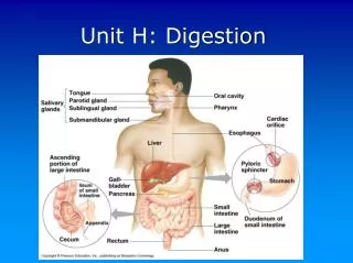
Digestion in the stomach: type & product (17) • Chemical breakdown of proteins begins and food is converted to chyme • Delivers chyme to the small intestine • Enzymaticallydigests proteins with pepsin
Function of goblet cells (18) • Epithelial lining is composed of Goblet cells that produce a coat of alkaline mucus • The mucous surface layer traps a bicarbonate-rich fluid beneath it
Gastric Pits (18) • Contain gastric glands that secrete gastric juice, mucus, and gastrin
Cells & their function in the stomach fundus and body (19) • Gastric glands of the fundus and body have a variety of secretory cells • Mucus neck cells secrete acid mucus • Parietal cells secrete HCl and intrinsic factor (Intrinsic factor is a glycoprotein necessary for the absorption of vitamin B12) • Chief cells produce pepsinogen
What is pepsinogen? • Activated to pepsin by HCl in the stomach • Pepsin (an enzyme) provides a positive feedback mechanism and functions to degrade food proteins to peptides (short polymers formed from the linking of amino acids)
Chief cells of the stomach secrete the digestive enzymes (pepsins) of the stomach
The stomach does not digest itself! (20) • The stomach produces a mucus lining secreted specialized cells which protects the stomach walls • These cells are continuously replaced to maintain the protective coating • Excessive secretions due to stress or smoking can form ulcers
Major functions of the stomach (21) • Holds ingested food • Degrades this food both physically and chemically • Delivers chyme to the small intestine • Enzymatically digests proteins with pepsin
Structural modification to increase surface area in the small intestine (22) • Plicae circulares - deep circular folds of the mucosa and submucosa of jejunum • Villi - fingerlike extensions of the mucosa • Microvilli - tiny projections of absorptive mucosal cells’ plasma membranes
Microvilli – brush borders of plasma membranes of intestinal cells
Small Intestine: Histology of the Wall The mucosal epithelium is made up of: • Absorptive cells, goblet and other cells • Cells of intestinal crypts secrete intestinal juice • Peyer’s patches are found in the submucosa of ileum • Brunner’s glands in the duodenum secrete alkaline mucus
Intestinal crypts – secretory glands in epithelial lining of small intestine
Brunner’s glands of duodenum Produce an alkaline secretion (containing bicarbonate) in order to: • protect the duodenum from the acidic content of chyme (which is introduced into the duodenum from the stomach); • provide an alkaline condition for the intestinal enzymes to be active, thus enabling absorption to take place; • lubricate the intestinal walls.
Digestive juice – composition & function (23) • Secreted by intestinal glands in response to distension or irritation of the mucosa • Slightly alkaline and isotonic with blood plasma • Largely water, enzyme-poor, but contains mucus