Download
1 / 19
190 likes | 249 Views
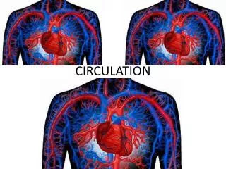
Circulation. The Presence of a Circulatory System:. Reduces the distance a substance must travel to enter or leave a cell. Uses “Blood” to carry these molecules to the cells.

E N D
The Presence of a Circulatory System: • Reduces the distance a substance must travel to enter or leave a cell. • Uses “Blood” to carry these molecules to the cells. O2 diffuses from the air in the lungs (or similar structures) across epithelium to the “blood” which carries it to all parts of the body. Once in the capillaries, O2 diffuses across epithelium again to the tissues. CO2, of course, moves in the opposite direction.
Circulatory systems • All animals have: • circulatory fluid = “blood” • tubes = blood vessels • muscular pump = heart open closed hemolymph blood
Open circulatory system • Invertebrates • insects, arthropods, mollusks • No separation between blood & interstitial fluid (Hemolymph)
Closed circulatory system closed system = higher pressures • Invertebrates • earthworms, squid, octopuses • Vertebrates • Blood confined to vessels & separate from interstitial fluid • 1 or more hearts • large vessels to smaller vessels • material diffuses between blood vessels & interstitial fluid
Vertebrate circulatory system • Adaptations in closed system • number of heart chambers differs 2 3 4 low O2to body low pressureto body high pressure & high O2to body What’s the adaptive value of a 4 chamber heart? 4 chamber heart is double pump = separates oxygen-rich & oxygen-poor blood; maintains high pressure
Evolution of vertebrate circulatory system fish amphibian reptiles birds & mammals 2chamber 3 chamber 3 chamber 4 chamber V A A A A A A A V V V V V
Evolution of 4-chambered heart • Selective forces • increase body size • protection from predation • bigger body = bigger stomach for herbivores • endothermy • can colonize more habitats • flight • decrease predation & increase prey capture • Effect of higher metabolic rate • greater need for energy, fuels, O2, waste removal • endothermic animals need 10x energy • need to deliver 10x fuel & O2 to cells convergentevolution
Vertebrate cardiovascular system • Chambered heart • atrium = receive blood • ventricle = pump blood out • Blood vessels • arteries= carry blood away from heart • arterioles • veins= return blood to heart • venules • capillaries = thin wall, exchange / diffusion • capillary beds = networks of capillaries
Exchange across capillary walls Pressure is greatest in the arteries, lowest in veins Lymphatic capillary Interstitial fluid flows back into capillaries due to osmosis • plasma proteins osmotic pressure in capillary Fluid & solutes flows out of capillaries to tissues due to blood pressure • “bulk flow” BP > OP BP < OP Interstitial fluid Blood flow 85% fluid returns to capillaries Capillary 15% fluid returns via lymph Arteriole Venule
Mammaliancirculation systemic pulmonary systemic What do bluevs.redareas represent?
Mammalian heart to neck & head& arms
SL AV AV Heart valves • 4 valves in the heart • flaps of connective tissue • prevent backflow • Atrioventricular (AV) valves • between atrium & ventricle • keeps blood from flowing back into atria when ventricles contract • “lub” • Semilunar valves • between ventricle & arteries • prevent backflow from arteries into ventricles while they are relaxing • “dub”
Lub-dub, lub-dub • Tempo is controlled by Sinoatrial node (SA node) • located at right atrium by superior vena cava • Contraction at SA node causes both atria to contract (lub) • Wave contraction passes down to Atrioventricular node (AV node) • Impulse is delayed by 0.1 sec (atria need to empty) • Causes ventricle to contract (dub) • Heart murmur • defect in valves causes hissing sound when stream of blood squirts backward through valve SL AV AV
Cardiac cycle 1 complete sequence of pumping heart contracts & pumps heart relaxes & chambers fill contraction phase systole ventricles pumps blood out relaxation phase diastole atria refill with blood 110 ____ 70 systolic ________ diastolic pump(peak pressure) _________________ fill(minimum pressure)
Measurement of blood pressure • High Blood Pressure (hypertension) • if top number (systolic pumping) > 150 • if bottom number (diastolicfilling) > 90
Blood: It’s more than just red stuff. Plasma: 55% The fluid portion of blood. Water accounts for over 90% Contains: Electrolytes- to maintain osmotic balance, buffer • Nutrients • Respiratory gases • Hormones CELLS! So, if blood is 55% plasma, the rest must be ….._________
Blood: Erythrocytes-also called Red Blood Cells (RBCs) So, what about those cells? • Transport oxygen (which binds to Hemoglobin) • Lack nuclei or mitochondria • Leukocytes- also called white blood cells (WBCs) • Function in defense and immunity • DO have nuclei & mitochondria Include: Basophils Actually spend most of their time in interstitial fluid and the lymphatic system. Lymphocytes Eosinophils Neutrophils
Clotting: a real fixer-upper… Platelets: • Clump & stick to the jagged edges of the cut. Provides immediate but temporary “plug” 2. Release clotting factors which catalyze the reaction: Fibrinogen Fibrin Fibrin aggregates to form the clot