Download
1 / 49
520 likes | 1.63k Views
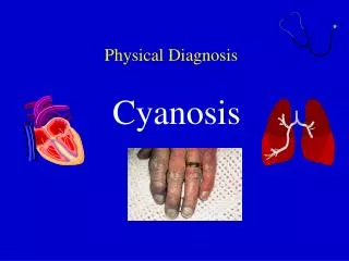
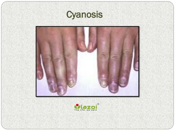
APPROACH TO CYANOSIS. Definition. Bluish discolouration of skin or mucous membrane caused by excess amounts of reduced hemoglobin or abnormal hemoglobin 4gm of reduced Hb in capillaries required for cyanosis to be apparent.

E N D
Definition Bluish discolouration of skin or mucous membrane caused by excess amounts of reduced hemoglobin or abnormal hemoglobin 4gm of reduced Hb in capillaries required for cyanosis to be apparent
Mechanismcaused by absolute increase in reduced Hb,higher the Hb – greater tendancy towards cyanosis
In severe anemia , greater systemic arterial desaturation required for cyanosis to be evident • In polycythemia even lesser systemic arterial oxygen saturation may result in clinical cyanosis • If fetal Hb is high, tissue hypoxia may occur even if cyanosis is mild( arterial PaO2 low)
CARDIOVASCULAR DUCT-INDEPENDENT MIXING LESIONS TGA, TAPVC,TA DUCT-DEPENDANT PBF TOF,EBSTEINS,TricusidAtre DUCT-Dependant SBF HLHS,IAA,CoA,Critical AS L-RSHUNT &PUL EDEMA SV States PRIMARY LUNG DISEASE AIRWAY OBSTRUCTION EXTRINSIC LUNG COMPRESSION PULMONARY AV MALFORMATION PPHN CNS DYSFUNCTION HEMATOLOGIC MISCELLANEOUS HYPOGLYCEMIA, METABOLICACIDOSIS SEPSIS, HYPOTHERMIA, SHOCK
MATERNAL HISTORY DIABETES- TTN,RDS,HYPOGLYCEMIA ASTHMA -TTN POLYHYDRAMNIOS - TEF PIH – IUGR,POLYCYTHEMIA,HYPOGLYCEMIA LABOUR& DELIVERY PROM –SEPSIS,PNEUMONIA CHORIOAMNIONITIS- SEPSIS C-SECTION- TTN,RDS,PPHN NEWBORN ONSET AT BIRTH- TTN,RDS,MAS,CDH, ONSET –HRS AFTER BIRTH- CCHD,aspiration,TEF
CLUES BASED ON ONSET 1st week > 1 week D TGATOF TGA Pulmonary AtresiaAdmixture lesions Tricuspid atresiaTAPVC EbsteinSV Critical PS DORV Truncus
Examined in neutral thermal enviornment Away from blue phototherapy lights Asses capillary refill time- <2 sec Barrel shaped chest –post term-MAS Bell shaped thorax – neurologic abnormalities Scaphoid abdomen-CDH Look for nasal flaring,grunting & retractions
Palpate brachial & femoral pulses BP- all four extrimities SBP-gradient quite specific for arch abnormality Not sensitive S2- split in 80% by 48 hours Differential cyanosis
CCHD in Newborns:Clues based on presentation Cyanosis No Resp Distress Cyanosis + Resp Distress Shock Differential cyanosis TGA DDPC TAPVC obstructed DDSC
CCHD in Newborns:Clues Based on S2 split S2 single normal fixed DDPC TGA DDSC Excludes Cardiac cause TAPVC
CCHD in newborns:Clues based on Murmurs • Murmurs have poor sensitivity( < 50%)
Pulse oximetry Standard of care for all infants with respiratory distress & cyanosis Accurate& reliable method of monitoring o2 saturation in infants noninvasively Pulse oximeter probes on R hand & lower extremity Aim for 02 saturation of 90-95% by pulse oximetry PPHN- suspected –aim for higher o2 saturation
HYPEROXIA TEST • ADMINISTER 100% O2 FOR 15 MINUTES • ASSES O2 OF UPPER LIMB LOWER LIMB • ABG - YES • TCMO - YES • PO - NO
HYPEROXIA TEST GIVE 100% O2 ASSES PO2 PO2>200PO2<150 NO CCHD LIKELY CCHD PASS FAIL 150-200 ?CCHD WITH PBF OR PPHN
PAO2 <50MM C X RAY CARDIOMEGALY NO CARDIOMEGALY PULMONARY VASCULARITY EBSTEINS PBF PUL EDEMA PBF TGA + IVS TAPVR With OBSTRN TA with PA orPS PA WITH IVS CRITICAL PS TOF & TOF + PA
Those with decreased PBF &normal or slightly increased heart size differentiated by there QRS axis on ECG & MURMUR + or – TA with PS or PA – SUPERIOR QRS AXIS ( 0 to-90) Critical PS & PA with IVS- 0 to 90 degree Differentiated by loud pul ejection murmur TOF & TOF with PA - QRS 90 to 180 Pulmonary continuous murmur Stenosis murmur
Prostaglandin (PGE1) Infusion Neonates –fail hyperoxia test High Signs& symptoms of CHD or likelihood Present in shock within 1st 3 wk of life of CHD PGE1 administration –open ductusarteriosus Depending on lesion - PBF or SBF or improves Intercirculatory mixing- improves hypoxemia &metabolic acidosis Neonate with shock or CHF in 1st few weeks of life - ductdependant SBF untilproven otherwise
PG sensitive lesions: Cyanosis + murmur or mild/no cyanosis + abnormal pulses • Can be withheld in a relatively stable child ( SaO2 > 70%; no acidosis) • Target SaO2 >80%; pO2 around 45-50, normal pH • Once diagnosis is confirmed, it is ideal to start PGE1 before transport. WORSENING AFTER PGE1 – obstruction to blood flow out of pulmonary veins
Always given as continous IV infusion. • Start at 0.05-0.1μg/kg/min, can be reduced to 0.005 - 0.01µg/kg/min once duct is opened( ^ SaO2) • Available as 500 μg vial • Trade name: Alpostin/Prostin • Cost: Rs 5000/- per ampoule • One vial will last 2-3 days for a 3Kg baby
Efficacy with age, less effective after 2 weeks of life, not effective after 4 weeks • Adverse reactions more common in premature& LBW infants • Apnea –typically in 1st few hrs ,tachycardia, bradycardia, fever, NEC, seizures, thrombocytopenia, • Continouscardiorespiratory monitoring
PGE1 – peripheral vasodilation – hypotension& cutaneous flushing Separate IV line should be secured Hypotension treated by 10-20ml/kg bolus of NS,RL,5%albumin RemeasureABG,reasses capillary refill& vitalsigns within 15 to 30 min of starting PGE1 infusion
Principles of managment Intialstabilisation – airway management reliable venous access – umblical vein Arterial line to monitor BP,acid-base,o2 Volume resucitation,inotropic support & correction of metabolic acidosis Blood glucose & sepsis workup-cyanosis+circcollpse
Non invasive delineation of anatomic defect- ECHO Evaluation & treatment of additional organ system-pulmonary,renal,hepatic&CNS Evaluation of additional congenital defects Genetic evaluation – if indicated Cardiac Cathetrisation Surgical managment
CCHD is an important differential diagnosis in neonate presenting with cyanosis after birth. • Clinical evaluation with CXR and Hyperoxia test excludes CHD in most cases. • Echocardiography recommended in all doubtful cases. • Prior stabilization and a monitored transport to tertiary center ensures a optimal pre-operative state. • Early intervention with very encouraging results is realistic for most forms of critical CHD in newborns
r CCHD derived from heterogeneous group of conditions May have pulmonicstenosis ,PAHor NL pulm pressure with out PS PBF may be NL,INCREASED or DECREASED Decreased PBF may be due to Pulmonicstenosis or PAH Anomalies where free mixing of systemic and venous blood occurs severity of cyanosis determined by pulmonary blood flow Thymus regresses very fast in cyanotic patients
This classification result in six sub group of cyanotic patients PS without VSD PS + large VSD Increased PBF with or without PAH(transposition physiology PAH withdecreased PBF(Eisenmengerphisiology) PAHdue to pul venous obstruction NL or MILD elevated pul pressure without PS
PS without VSD (cyanosis due to R toL shunt at atrial level& cardiomegaly) Triad-cyanosis,cardiomegaly,ischemic lung fields on Cxray Dominant a in JVP Cardiomegaly Parasternalimpulse Widely split 2nd HS p2 late &soft 3rd &4th HS Pulm ejection systolic murmur,TR murmur Severe or critical pure PS with failing RV,Ebstein’s
PS with VSD (Fallot’s physiology) Prominent a wave in jvp NL heart size Mild parasternal impulse Systolic thrill uncommon Single 2nd sound(widely split with inaudible pulmonic sound) ESM Clear diastolic period Ischemic lungs in xray without cardiomegaly
D/D of fallots physiology Fallot’stetrology(commonest >2yrs ) TGA TRICUSPID ATRESIA SV DORVcorrectedTGA AVCD If no rvh on ecg;posssibilities-rvhypoplastic &small,rvabsent,PA not connected to RV Tricuspid atresia,hypoplasticRV with or without straddling TV, & single ventricle
Increased pulmonary blood flow with or without PAH(transposition physiology) Symptomatic in Neonate Cyanosis-mild to severe Failure to thrive& gain weight CCF Cardiomegaly –in2-3 wks of life 2nd sound single,s3 gallop,insignificant systolic murmur Cardiomegaly with Incrspulmvasculatureonxray& thymicshadow absent
Anomalies with cyanosis and increased PBF TGA DORV without PS TA with Incrs PBF (largeVSD+ NO PS) Persistent Truncus SV without PS TAPVC
Cyanosis with PAH and Diminished PBF (eisenmenger- defnd as nonreactive PAH resulting in a R to L shunt at atrial,ventricular or great artery level) Characteristics H/O frequent chest infections in infancy Cyanosis present from birth and appear late Jvp-prominent a wave No cardiomegaly or thrill(except when shunt at atrial level) No PSH Constant EC of PAH
2nd sound is palpable,pulmonary component accentuated Systolic murmur in pulmonary area is insignificant or absent Pulmonary and/or TR murmurs may be present
Patients with PAH due to Pulmonary Venous hypertension(HLHS&TAPVC with obstruction) Generally present in neonatal period Severe cyanosis,CHF,S3 gallop,nocardiomegaly,absence of significant murmurs, Cxray –NL sized heart with severe PVH- causing GROUND GLASS appearance 2d echo- mitralatresia,aorticatresia,pulmonary venous obstruction, hypoplastic LV
Cyanosis without PS & PA pressure normal Heterogeneous group of anomalies TAPVC- features of 2 ASD but with cyanosis,figure of 8 Single atrium-mostly associated with polysplenia SVC entering LA Pulmonary AV fistula
Abnormal rhythm& CHD • L- TGA - Heart Block ; SVT • Ebstein - SVT