Download
1 / 36
360 likes | 373 Views
Delve into the intricate layers and functions of the skin, from the epidermis to the dermis, and learn about skin color, burns, and appendages like hair, nails, and glands. Unravel the complexities of this vital organ!

E N D
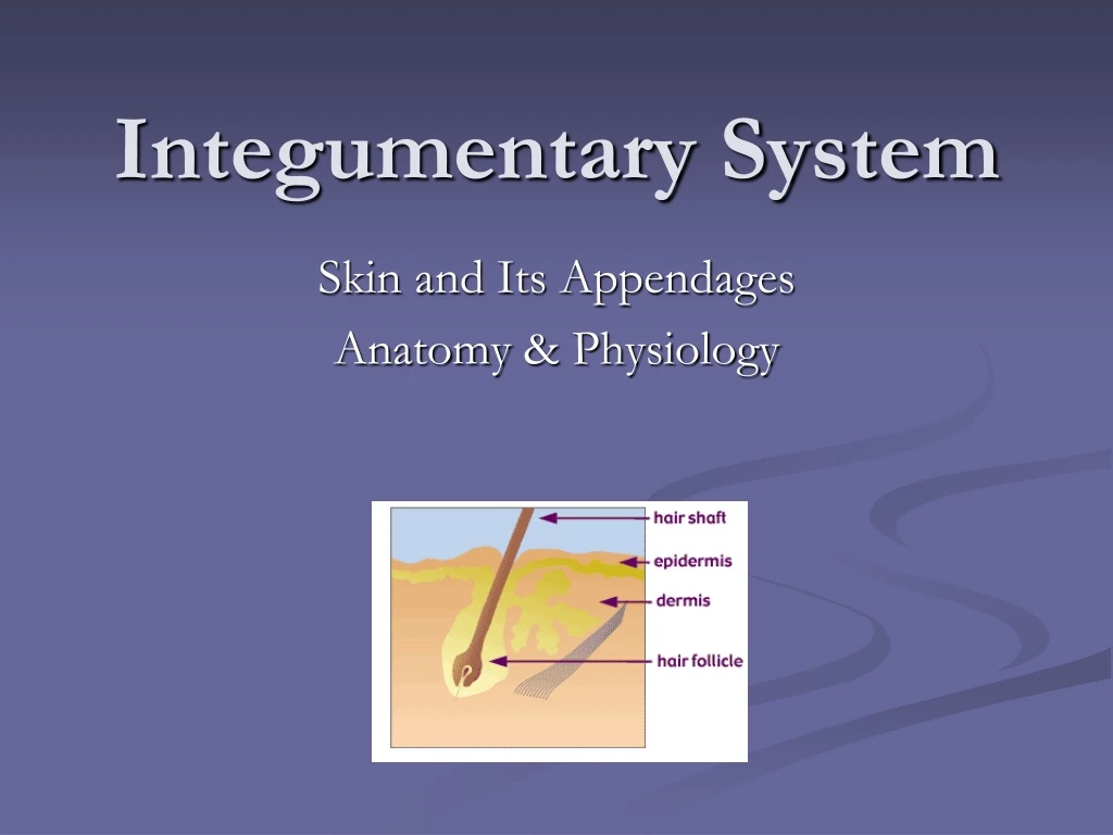
Integumentary System Skin and Its Appendages Anatomy & Physiology
Skin or Integument • Largest organ in the body • Integumentary System: denotes the skin and its appendages
Layers of the skin • Epidermis: Outer, thinner layer • Dermis: Thicker layer, connective tissue • Hypodermis: Subcutaneous layer, superficial fascia
Thick Skin • Refers to epidermal layer only • Found: palms of hands, soles of feet, fingertips • Each of the 5 layers present • Dermal papillae: fingerprints • No hair
Cell Types of Epidermis • Keratinocytes: contain keratin, make up 90% of epidermal cells • Melanocytes: contribute color to skin • Langerhanscells: immunological reactions in skin
Cell Layers of Epidermis • Stratum corneum • Stratum lucidum • Stratum granulosum • Stratum spinosum • Stratum basale
Stratum corneum (horny layer) • Flat thin squamous cells • Surface cells dead & continually being shed • Cytoplasm in cells replaced by keratin • Desmosomes hold cells together • Barrier layer of the skin
Stratum lucidum (clear layer) • Nuclei absent • Cells contain eleidin which will be transformed to keratin • Blocks water penetration or loss • Absent from thin skin
Stratum granulosum (granular layer) • Keratinization begins • 2-4 layers deep • Maybe absent in thin skin • Cells filled with granules called keratohyalin
Stratum spinosum (spiny layer) • 8-10 layers with prominent desmosomes which appear spiny under a microscope • Cells rich in RNA
Stratum basale (base layer) • Single layer of columnar cells • Only cells which undergo mitosis • Cells migrate from basal layer thru the outer layers
Dermal-Epidermal Junction • Contains basement membrane • Also contains a polysaccharide gel that “glues” 2 layers together
Dermis • Thin papillary layer & thick reticular layer • Thickest on soles & palms • Thinnest on eyelids & penis • Mechanical strength of skin
Papillary layer • Forms the bumps, dermal papillae which project into epidermis • Allows us to grip surfaces & creates fingerprints
Reticular layer • More dense collagen & elastic fibers • Serves as point of attachment for muscle fibers • Skeletal muscle: muscles of facial expression • Smooth muscle: arrectorpili muscles on hair follicles
Skin Color • Determined by quantity of melanin in cells of epidermis • All races have about the same number of melanocytes but differ in amount of melanin produced • Sun can increase melanin production
Functions of skin • Protection • Sensation • Movement without injury • Vitamin D production • Excretion • Immunity • Temperature regulation
Heat Loss • Evaporation • Radiation • Conduction • Convection
Burns • Predict body surface area to determine how much fluid to replace: • Rule of palms (1%) • Rule of nines
First degree burn • Involves only the epidermis • No blistering or scarring • Sunburn • Reddening of the skin, mild discomfort
Second degree burn • Involves epidermis & dermis • Blistering, pain, swelling • May scar
Third degree burn • Destruction of epidermis & dermis, may involve underlying tissue • Severe scarring
Appendages of the skin • Hair • Nails • Skin glands
Hair • Lanugo hair: fine hair covering fetus • Vellus hair: replacement for lanugo hair, first appears on scalp, eyelids, eyebrows • Terminal hair: Coarse hair that replaces vellus hair-axillary, pubic, beard, chest & hair on arms & legs in men
Hair follicle • Stratum germinativum develops into follicle's inner layer and forms the germinal matrix • Small mound of dermis protruding into germinal matrix is the papilla (contains blood capillaries)
Parts of the Nail Matrix- the thickened, proximal area of the nail that is responsible for growth Bed- the hard translucent visible part of the nail Root- the point of attachment under the skin Cuticle- the layer of skin that prevents dirt and bacteria from getting into the nail bed Free Body- the end of the nail that is not connected to the skin
Glands • Sweat or sudoriferous glands • Eccrine sweat gland • Apocrine sweat glands • Sebaceous glands • Ceruminous glands
Eccrine sweat glands • Most numerous • Over most of the body • Secretory portion located in the subcutaneous tissue • Simple coiled tubular gland
Apocrine Sweat glands • Found in armpit, areola of breast, around the anus • Large than eccrine • Connected with hair follicles
Sebaceous glands • Secrete sebum into each follicle
Ceruminous glands • Modification of apocrine sweat glands • Open into ears • Produce cerumen
Image Citations • Slide 1: cross section of skin, 7/12/06, http://vilenski.org/science/humanbody/hb_html/skin.html • Slide 3: Anthony’s Textbook of Anatomy & Physiology, Seventeenth Edition by Thibodeau & Patton, Chapter 6. • Slide 5: Thick skin, 7/30/06, http://erl.pathology.iupui.edu/HISTO/LABE151.HTM • Slide 6: Melanocytes, 7/30/06, http://www.lab.anhb.uwa.edu.au/mb140/CorePages/Integumentary/Integum.htm • Slide 7: Thick skin trichrome, 7/30/06, http://www.lab.anhb.uwa.edu.au/mb140/CorePages/Integumentary/Integum.htm • Slide 8: Slide 43, Thick skin, 7/30/06, http://w3.ouhsc.edu/histology/Glass%20slides/43_09.jpg • Slide 9: Stratum lucidum human foot, 7/30/06, http://oregonstate.edu/~hanba/Projector%20Slides/Projector%20Slides/Skin%20Stratum%20Lucidum%20Human%20Foot-2.jpg • Slide 10: Stratum granulosum, 7/30/06, http://anatomy.iupui.edu/courses/histo_D502/D502f04/Labs.f04/Lab14/s31.100x.i3.jpg • Slide 11: Stratum spinosum, 7/30/06, http://anatomy.iupui.edu/courses/histo_D502/D502f04/Labs.f04/Lab14/s31.100x.i2.jpg • Slide 12: Stratum basale, 7/30/06, http://online-media.uni-marburg.de/histologie/introhis/HIS/skin/skin06.gif • Slide 14: Dermis, 7/30/06, http://sprojects.mmi.mcgill.ca/dermatology/dermis.htm • Slide 15: Dermis, 7/30/06, http://neuromedia.neurobio.ucla.edu/campbell/skin/wp_images/7%20dermis.jpg • Slide 16: 7/30/06, http://www.potterleague.org/Potter_Kids_Final/pet_body_lang.htm
Image Citations • Slide 20: Wallace’s rule of nines, 7/30/06, http://www.sunmed.org/burns.html • Slide 21: First degree burn, 7/30/06, http://www.grossmanburncenter.com/orig-site/web/care/causes.htm • Slide 22: Burn symptoms, 7/30/06, http://www.maggiessecret.com.au/burns-scalds.aspx • Slide 23: Third degree burn, 7/30/06, http://www.grossmanburncenter.com/orig-site/web/care/causes.htm • Slide 26: Thibodeau & Patton, Anthony’s Textbook of Anatomy & Physiology, Seventeenth Edition. • Slide 27: Thin Skin, 7/30/06, http://biology.clc.uc.edu/fankhauser/Labs/Anatomy_&_Physiology/A&P201/Integumentary/hair_follicle_100x_PA112040labeled.JPG • Slide 28: Sebaceous gland and shaft of hair follicle, 7/30/06, http://www.keele.ac.uk/depts/ms/resources/anatomy/histologyimages/t146.html • Slide 29: “Structure of nails”, Thibodeau & Patton, Anthony’s Textbook of Anatomy & Physiology, Seventeenth Edition. • Slide 31: “Skin Glands”, Thibodeau & Patton, Anthony’s Textbook of Anatomy & Physiology, Seventeenth Edition. • Slide 32: Dermis (Apocrine sweat glands), 7/30/06, http://www3.umdnj.edu/histsweb/lab11/lab11apocrine.html


