1 / 1

Nigrosome 1 in T2*w in vivo 7T MRI: all HC and PD subjects

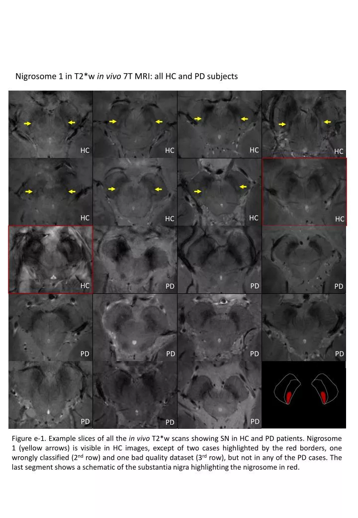
HC. HC. HC. HC. Nigrosome 1 in T2*w in vivo 7T MRI: all HC and PD subjects. HC. HC. HC. HC. HC. PD. PD. PD. PD. PD. PD. PD. PD. PD. PD.

lloyd
Download Presentation

Nigrosome 1 in T2*w in vivo 7T MRI: all HC and PD subjects

An Image/Link below is provided (as is) to download presentation Download Policy: Content on the Website is provided to you AS IS for your information and personal use and may not be sold / licensed / shared on other websites without getting consent from its author. Content is provided to you AS IS for your information and personal use only. Download presentation by click this link. While downloading, if for some reason you are not able to download a presentation, the publisher may have deleted the file from their server. During download, if you can't get a presentation, the file might be deleted by the publisher.

E N D

Presentation Transcript


  1. HC HC HC HC Nigrosome 1 in T2*w in vivo7T MRI: all HC and PD subjects HC HC HC HC HC PD PD PD PD PD PD PD PD PD PD Figure e-1. Example slices of all the in vivo T2*w scans showing SN in HC and PD patients. Nigrosome 1 (yellow arrows) is visible in HC images, except of two cases highlighted by the red borders, one wrongly classified (2nd row) and one bad quality dataset (3rd row), but not in any of the PD cases. The last segment shows a schematic of the substantianigra highlighting the nigrosome in red.

More Related