Download
1 / 112
1.18k likes | 1.35k Views
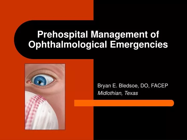
Prehospital Management of Ophthalmological Emergencies. Bryan E. Bledsoe, DO, FACEP Midlothian, Texas. Ophthalmological Emergencies. The eyes are the windows to the soul. Eye-injuries can be life-changing events and EMS personnel should provide the best care possible to save a person’s sight.

E N D
Prehospital Management of Ophthalmological Emergencies Bryan E. Bledsoe, DO, FACEP Midlothian, Texas
Ophthalmological Emergencies • The eyes are the windows to the soul. • Eye-injuries can be life-changing events and EMS personnel should provide the best care possible to save a person’s sight.
Ophthalmological Emergencies • Anatomy and Physiology • Assessment • Medical Conditions • Traumatic Conditions • Prehospital Management
Anatomy and Physiology • External Anatomy • Boney Anatomy • Associated Structures • Extra-ocular Muscles • Eye Anatomy • Chambers • Retina • Neurological Anatomy
Assessment • History • Physical Examination • Visual Acuity • External Eye • Confrontation/Visual Fields • Pupils • Ocular Motility • Anterior Segment • Fundus* • IOP* * May not be appropriate for EMS except in special circumstances
History • Onset (Slow versus rapid) • Monocular versus Binocular • Antecedent activities (hammering) • Past visual acuity (need for glasses) • Unusual signs/symptoms • Other medical conditions
Medical Conditions • Stye (External Hordeolum) • Chalazion (Internal Hordeolum)
Stye (External Hordeolum) • Staph infection of oil gland associated with an eyelash. • Located at lash line and has appearance of small pustule.
Stye (External Hordeolum) • Treated with warm soaks and topical ophthalmic antibiotics.
Chalazion (Internal Hordeolum) • Acute or chronic inflammation secondary to blockage of one of the meibomian oil glands in the tarsal plate. • Red, tender lump in the lid or at the lid margin
Chalazion (Internal Hordeolum) • Approximately 50 glands on the upper lid and 25 on the lower lid. • Glands serve to keep the eye moist by spreading sheet of oil across the eye with blinking.
Chalazion (Internal Hordeolum) • Treatment: • Warm compresses 3-4 times a day. • Topical ophthalmic antibiotics. • Oral antibiotics. • Ophthalmology referral.
Conjunctiva • Bacterial Conjunctivitis • Viral Conjunctivitis • Allergic Conjunctivitis • Neonatal Conjunctivitis • Pterygium
Bacterial Conjunctivitis • Irritation of the conjunctiva and purulent drainage. • Cornea is clear. • Commonly referred to as “pink eye”.
Bacterial Conjunctivitis • Treatment: • Topical antibiotics. • Analgesia
Allergic Conjunctivitis • Inflammation of the conjunctiva due to allergens in the environment. • Prominent redness and itching. • Cornea clear.
Allergic Conjunctivitis • Treatment: • Artificial tears. • Topical antihistamines/ decongestants.
Allergic Conjunctivitis • Treatment: • Severe cases may require ophthalmic steroids.
Neonatal Conjunctivitis • Conjunctivitis (Neonatal) • Caused by Neisseria gonorrhoeae, Chlamydia, or Herpes virus. • Infant must be evaluated to exclude systematic infection.
Pterygium • Raised web-shaped growth of the conjunctiva. • More common in sunny and tropical climates. • Can invade the cornea.
Pterygium • Sometimes it spontaneously resolves. • Surgery necessary in other cases.
Corneal Disease • HSV Keratitis • Herpes Zoster Ophthalmicus • Corneal Ulcers
HSV Keratitis • Can affect eyelids, conjunctiva and cornea. • Typical dendritic appearance can be seen in the cornea.
HSV Keratitis • Caused by Herpes Simplex Virus. • Can cause permanent corneal scarring.
Herpes Zoster Ophthalmicus • Shingles in the distribution of the trigeminal nerve. • Caused by reactivation of the Herpes zoster virus.
Corneal Ulcers • Serious infection involving multiple layers of the cornea. • Caused by entry of infectious agents through breaks in the epithelial border.
Corneal Ulcers • Patient usually has: • Painful red eye • Tearing • Photophobia • Treatment: • Topical antibiotics • Cyloplegics
Cellulitis • Preseptal (Periorbital) Cellulitis • Postseptal (Orbital) Cellulitis
Periorbital Cellulitis • Cellulitis that has not breached the orbital septum. • Eyelids edematous, warm and red. • Eye not involved. • Staph., Strep., and viruses common cause.