Download
1 / 54
550 likes | 806 Views
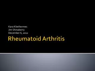
RHEUMATOID ARTHRITIS (RA). RHEUMATOID ARTHRITIS ( RA ) Gergely Péter dr. Definition: Chro ni c destru ctive diseases characterized by joint inflammation with pain and swelling. In a considerable proportion of patients, the arthritis is progressive, resulting in joint destruction and

E N D
RHEUMATOID ARTHRITIS (RA) RHEUMATOID ARTHRITIS (RA) Gergely Péter dr Definition: Chronic destructivediseases characterized by joint inflammation with pain and swelling. In a considerable proportion of patients, the arthritis is progressive, resulting in joint destruction and ultimately incapacitation and increased mortality. Relatively common, prevalence: 0.3-1.5 %, themale:femaleratiocca. 1:3. Typical case: womanaged 30-40 years with polyarthritis and early joint deformities.
Pathogenesis of RA Endogenous factors Exogenous factors MHC genes, hormon milieu) cross-reacting antigenes, bacteria, viruses Synovial vasculitis Adhesion molecule expression cellular infiltration Macrophages, T cells, B cells, granulocytes Cytokines (TNF-a, IL-1, IL-6), RF, free-radicals, enzymes Synovial proliferation, angiogenesis, chondrocyte-, osteoclast- activation Pannus, cartilage destruction, bone resorption Pathogenesis of RA
Cytokineinteractions Cytokine interactions Cytokinek interakciói
Classification criteria of RA (ARA, 1987) • Morning stiffness – for at least 1 hr and present for atleast 6 weeks • Swelling of 3 or more joints for at least 6 weeks • Swelling of wrist, metacarpophalangeal (MCP) orproximal interphalangeal (PIP) joints for at least 6weeks • Symmetric joint swelling • Typical radiologic changes in hands (erosions orunequivocal bony decalcification) • Rheumatoid nodules • Serum rheumatoid factor (RF) positivity • Diagnosis is made by the presence of 4 or more criteria
Differential diagnosis of polyarthritis • RA should be differentiated from: • Other autoimmune diseases (SLE, primary Sjögren’s syndrome, MCTD, PM/DM,PSS, PAN, gian cell vasculitis, polymyalgia rheumatica, adult onset Still’sdisease) • Viral diseases (parvovirus B19 infection, rubella, hepatitis B &Cinfection) • Bacterial infections (tbc, rheumatic fever, Jaccoud’s arthritis, septicendocarditis, mycoplasma arthritis) • Seronegative spondylarthritides(erosive psoriatic arthitis, reactivearthritis, enteropathic arthritis) • Paraneoplastic arthritis • Other diseases (e.g. hyperthrophic osteoarthropathy,erythema nodosum, agammaglobulinemia, acromegaly, diabetes mellitus) • Other rheumatic diseases (chronic gout, inflamed erosive osteoarthritis) Differential diagnosis of polyarthritis
Signs of early RA (=undifferentiated arthritis) In the early stage (within the first 3-6 months) (ARA) classification criteria cannot be used. The patient should be referred to a rheumatologist, if • the patient has 3 or more swollen joints • the metacarpophalangeal (MCP) and/or metatarsophalangeal (MTP) joints are involved; the squeeze test is positive • morning stiffness is 30 min or more.
Squeeze test Squeeze test
Joint involvement in RA The most specific sign of RA is arthritis. It is progressive and deforming in the majority (2/3) of cases (= erosive polyarthritis)
RA early stage RA early stage
Early assymmetric RA Early assymmetric RA
PIP joint involvement in RA PIP joint involvement in RA
RA: swan neck deformity RA: swan neck deformity
RA: ulnar deviation RA: ulnar deviation
Ulnar deviation in RA with severe atrophy of interossealmuscles Ulnar deviation in RA with severe atrophy of interosseal muscles
RA: Boutonnière deformity RA: Boutonnière deformity
RA: arthritis mutilans RA: arthritis mutilans
Involvement of joints of feet in RA Involvement of joints of feet in RA
Severe destruction of ankles in RA Severe destruction of ankles in RA
Periarticular osteoporosis (decalcification) Periarticular osteoporosis (decalcification)
Erosions and sclerosis (in late stage) Erosions and sclerosis (in late stage)
Erosion in RA Erosion in RA
Early erosions (MRI) Early erosions (MRI)
Scinti-graphy ofthe hands Scinti- graphy of the hands
Baker’s cyst Baker’s cyst
Bursitis in the shoulder Bursitis in the shoulder
Bursitis and rheumatoid nodule Bursitis and rheumatoid nodule
Rheumatoid nodules Rheumatoid nodules
Atlantoaxialsubluxation Atlantoaxial subluxation
RA – end stage RA – end stage
Extraarticular manifestations of RA • rheumatoid nodules – subcutaneous - in internal organs (lung, aortic valve) • pleuritis/pericarditis • fibrotizing alveolitis • Felty’s syndrome • vasculitis • amyloidosis
Systemic manifestations of RA: pulmonary fibrosis Systemic manifestations of RA: pulmonary fibrosis
Interstitial pneumonitis in RA Interstitial pneumonitis in RA
Systemic manifestations of RA: Caplan’s syndrome Systemic manifestations of RA: Caplan’s syndrome
Rheumatoid nodules in the lungs Rheumatoid nodules in the lungs
Episcleritis in RA Episcleritis in RA
Scleritis in RA Scleritis in RA
Scleromalacia perforans Scleromalacia perforans
Vasculitis in RA Vasculitis in RA
Vasculitis in RA Vasculitis in RA
Leg ulcers in Felty’s syndrome Leg ulcers in Felty’s syndrome
Large granular lymphocytes in Felty’s syndrome Large granular lymphocytes in Felty’s syndrome
Disease modifying antirheumatic drugs (DMARD) Disease modifying antirheumatic drugs (DMARD): Drug Adverse effects Dose gold (i.m.)dermatitis, stomatitis, 25-50 mg /2-4 proteinuria, enterocolitis, weeks thrombocytopenia gold (p.o.) less frequently used, brecause of lower tolerability chloroquine (hydroxy- retinopathia, pigment- 250 mg/day chloroquine) anomalies Regular ophthalmology check is required d-penicillamine proteinuria, myasthenia, 125-750 mg/day stomatitis Owing to low tolerability it is not used any more azathioprine hepatitis, bone marrow depression 50-150 mg/day Scarcely given in RA methotrexatehepatotoxicity, pulmonary fibrosis, 7,5-25 (MTX) bone marrow depression mg/week most frequently used therapy
sulfasalazine sulfasalazine nausea, vomiting 1,5-2 g/day diarrhea, bone marrow depression cyclosporine A nephrotoxicity, tremor 1,5-4 mg/kg/day creatinineand blood pressure should be checked regularly leflunomidehepatotoxicity, GI 10-20 mg/day complaints TNF-blockers: local reaction, autoimmune disease (SLE, SM) (etanercept, infection (tbc) infliximab, and abatacept) etanercept: 25 mg 2x weekly s.c. infliximab: 3 mg/kg every 8 week i.v. Other: anakinra (IL-1 blocker) rituximab (anti-CD20 antibody) abatacept (T cell activation blocker antibody)
Diseases related to RA: • Diseases related to RA: • Juvenile forms (= juvenile RA, juvenile idiopathicarthritis (JIA) • Subgroups: • systemic (Still’s disease) • pauciarticular (<4 joints) • polyarticular (similar to adult RA) • 2) Seronegative (RF negative) forms (seronegative • spondarthropathies = SNSA) • Ankylosing spondylarthritis (Mo Bechterew) • Psoriatic arthritis • Reiter’s disease - postinfectious arthritis • Enteropathic arthritis
Classification criteria of JIA (ARA, 1982) 1. Persistent arthritis of at least 6 weeks duration in one or more joints 2. Exclusion of other causes of arthritis (in particular): a. other systemic autoimmune diseses (SLE, rheumatic fevers, vasculitis, PSS, SS, MCTD, Behçet’s syndrome, PM/DM, SPA, Reiter’s syndroma, psoriatic arthritis) b. Infectious arthritis c. Inflammatory bowel diseases d. Neoplasms (e.g. leukaemia) e. Nonrhematic conditions f. Hematologicdiseases g. Psychogenic arthralgia h. Other (sarcoidosis, hyperthrophicosteoarthropathy, villonodular synovitis, chronic aktive hepatitis, familial Mediterraneanfever)
Child with advanced polyarticular JIA Child with advanced polyarticular JIA
Micrognathia in JIA Micrognathia in JIA
Typical skin rash in Still’s disease Typical skin rash inStill’s disease
Inflamed joints with diffuse edema in SNSA (‘sausage-like’ fingers) Inflamed joints with diffuse edema in SNSA (‘sausage-like’ fingers)