Download
1 / 55
550 likes | 572 Views
This article discusses the key roles of nucleotides in various cellular processes, including RNA and DNA activation, co-enzymes components, biosynthetic processes, metabolic regulation, and energy currency. It also covers the nomenclature, de novo synthesis pathways, re-utilization pathways, and metabolic diseases associated with purine metabolism. The regulation of nucleotides, structures of common purine bases, and salvage pathways for the re-utilization of purines are also explained. Additionally, the article explores the biosynthesis of purines, the major regulatory step, and the degradation of purines to urate.

E N D
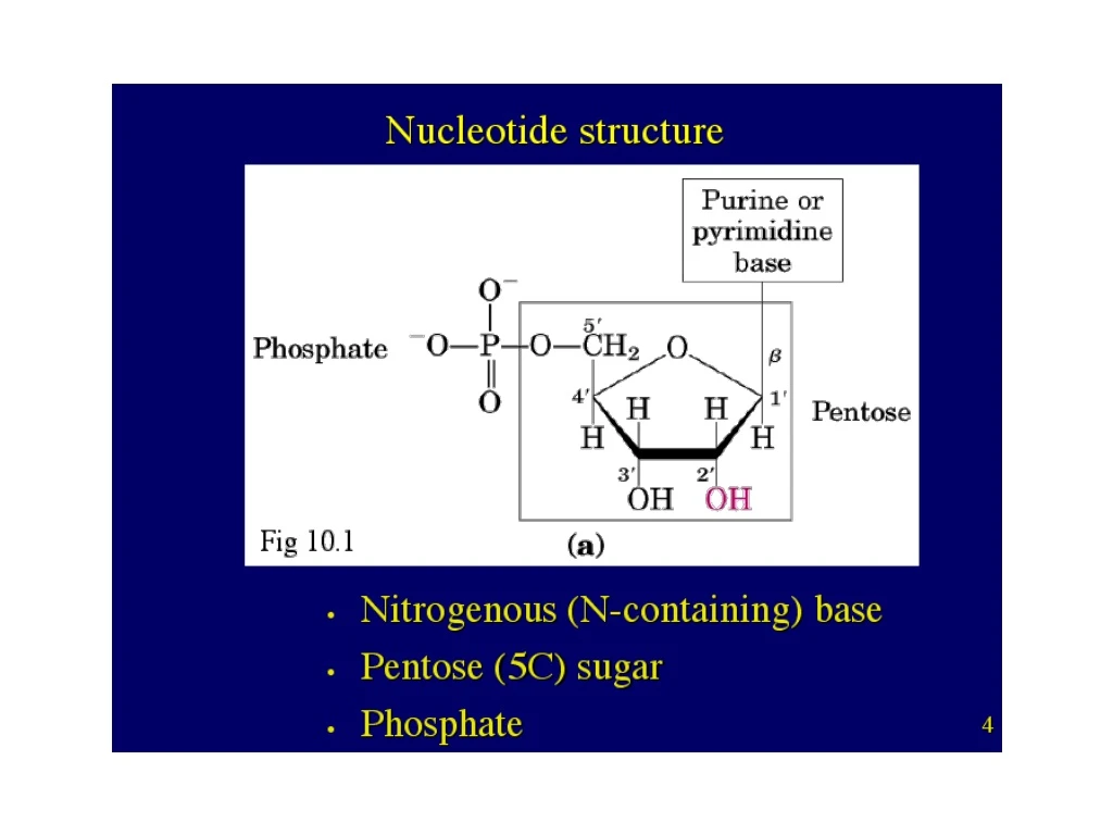
Nucleotides play key roles in many, many cellular processes 1. Activated precursors of RNA and DNA 2. Adenine nucleotides are components of three major co-enzymes, NAD, FAD, and CoA 3. Nucleotide derivatives are activated intermediates in biosynthetic processes (UDP-glucose, SAM) 4. Serve as metabolic regulators (for example cAMP and the activation of cell signaling. 5. Serve as major currency of energy in all cells (ATP and GTP). 6. Many metabolic diseases have their etiology in nucleotide metabolism.
Purine metabolism (Overview) 1. Nomenclature/nucleotide structure 2. De novo synthesis pathways 3. Re-utilization pathways 4. Metabolic diseases of purine Metabolism(Gout, Lesch-Nyham, SCID)
Thymidylate synthase dUMP dTMP N5,N10-methylene- tetrahydrofolate Dihydrofolate Thymidylate Synthase Tetrahydrofolate Why go through to the trouble to convert Uracil to Thymine? reduced oxidized NADPH Dihydrofolate reductase Serine transhydroxymethylase NADP+
The nomenclature of purines and pyrimidines depends on their linkage to a pentose Cytosine Cytidine Cytidine Monophosphate Nucleoside* Base Base Nucleotide Base (P04 ester) * when the base is purine, then the nucleoside ends in OSINE (AdenOSINE, GuanOSINE, InOSINE) when the base is pyrimidine, then the nucleoside ends in IDINE (UrIDINE, CytIDINE, ThymIDINE)
CMP + ATP XDP + YTP CDP + ADP XTP + YDP The active forms of nucleotides in biosynthesis and energy conversions are di and triphosphates Nucleoside Monophosphate Kinase Nucleoside Diphosphate Kinase
RIBONUCLEOTIDE REDUCTASE 1. Complex enzymatic reaction whereby electrons are transferred from NADPH through a series of sufhydryl groups at the catalytic site of Ribonucleotide Reductase. 2. Active site of RR contains thioredoxin, a 12 kD protein with two exposed cysteines, which become oxidized. 3. This ultimately allows for the reduction of ribose. REGULATION 1. Based on the response to cellular need for dATPs. NADPH NADP+ dATP is general inhibitor ATP is a general activator
Nucleotides are linked by 5’ to 3’ phosphodiester bonds to generate DNA and RNA
Structures of Common Purine Bases. H= 6 oxy purine X= 2,6 dioxy purine A= 6 amino purine G= 2 amino, 6-oxy purine
Structures of Common Purine Bases. H= 6 oxy purine X= 2,6 dioxy purine A= 6 amino purine G= 2 amino, 6-oxy purine
Structures of Common Purine Bases. (N source) Aspartate (N source) Glutamine The common mechanistic them for the conversion of A and G is the conversion of a carbonyl oxygen to an amino group
There are two basic mechanisms to generate purines and pyrimidines 1. DE NOVO BIOSYNTHETIC PATHWAYS (building the bases from simple building blocks) 2. SALVAGE PATHWAYS (the reutilization of bases from dietary or catabolic sources)
The biosynthesis of purine (A and G) begins with the synthesis of the ribose-phosphate Pentose phosphate pathway Ribose phosphate pyrophosphoKINASE
Glutamate Glutamine PRPP The major regulatory step in purine biosynthesis is the conversion of PRPP to 5-Phosphoribosyl-1-amine * PPi Amidophosphoribosyl transferase Amidophosphoribosyl transferase is a important regulatory enzyme in purine biosynthesis. It is strongly inhibited by the end products IMP, AMP, and GMP. This type of inhibition is called FEEDBACK INHIBITION.
Several amino acids are utilized in purine biosynthesis, IMP is the precursor for both AMP and GMP, the base is also called hypoxanthine
Structures of Common Purine Bases. (N source) Aspartate (N source) Glutamine The common mechanistic them for the conversion of A and G is the conversion of a carbonyl oxygen to an amino group
Purines:where do the atoms come from? Purine intermediates include: 1. Glycine 2. 1 C units of 5,10 mTHF 3. Glutamine 4. Asparate
Ribose 5-phosphate PRPP The regulation of purine biosynthesis is a classic example of negative feedback Inhibited by AMP AMP Phosphoribosyl amine IMP GMP Inhibited by IMP, AMP, and GMP Inhibited by GMP
Nucleotidase Phosphorylase Cytosine Cytidine Cytidine Monophosphate Nucleoside* Base Base Nucleotide Base (P04 ester)
PRPP + Adenine Adenylate Salvage pathways for the re-utilization of purines; There are 2 salvage enzymes with different specificities; 1. Adenine phosphoribosyl transferase 2. Hypoxanthine-guanine phosphoribosyl transferase + PPi + Guanine A-PRT HG-PRT PRPP + Guanine Guanylate
What happens in gout? Inhibited by AMP AMP Ribose 5-phosphate PRPP Phosphoribosyl amine IMP GMP Inhibited by IMP, AMP, and GMP Inhibited by GMP 1. Negative regulation of PRPP Synthatase & PRPP Amidotransferase is lost 2. PRPP levels are increased because of defects in salvage pathways Therefore, there is net increase in biosynthetic/degradation pathways!!
“By Royal Authority” by George Cruickshank. 19th century.
David Wells New York Yankees
Purines in humans are degraded to Urate Important points: 1. Nucleotides are constantly undergoing turnover! 2. There are many enzymes involved; Nucleotidases Nucleoside phosphorylases Deaminases Xanthine oxidases 3. the final common intermediate in humans is Urate, which is excreted. 4. there are several metabolic disorders resulting from defects in purine catabolism.
Serum Uric Acid Levels(mg/dl) Incidence of Gout (% of cases) >9.0 ~10% 7-9 0.5-3.5% <7.0 0.1% Hypoxanthine Guanine xanthine oxidase Xanthine Urate xanthine oxidase GOUT (Gouty Arthritis): A defect of purine metabolism Allopurinol: a. decrease urate b. increase xanthine & hypoxanthine c. decrease PRPP
AMP H20 Nucleotidase Pi Adenosine H20 Adenine deaminase* NH3 Inosine Hypoxanthine SCID-Severe Combined Immunodeficiency Syndrome Autosomal recessive disorder Mutations in ADA Infants subject to bacterial, candidiasis, viral, protazoal infections Both T and B cells are significantly Reduced (dATP is toxic) 1995-AdV expressing ADA was sucessfullly employed as gene therapy strategy
Disorders of Purine Metabolism: Disorder Defect Comments Gout PRPP synthase/ Hyperuricemia HGPRT Lesch Nyhan lack of HGPRT Hyperuricemia syndrome SCID ADA high levels of dAMP von Gierke’s disease glucose -6-PTPase Hyperuricemia
Structure of Pyrimidines C= 2 oxy, 4 amino pyrimidine T= 2,4 dioxy 5-methyl pyrmidine U= 2,4 dioxy pyrimidine O= 2,4 dioxy 6 carboxy pyrimidine
Pyrimidine biosynthesis: (occurs in cytosol) Pyrimidine biosynthesis begins with the assembly of the ring, then linked To ribose phosphate. Precursors are Glutamine (NH2), Bicarbonate (C) , and ATP (PO4). Q. Why is it advantageous to generate carbamoyl phosphate in the cytosol rather than the mitochondria?
committed step in pyrimidine biosynthesis Carbamoyl phosphate synthase II, ATCase, and Dihydrooratase are linked in a single 240 kD polypeptide chain. The enzyme is sometimes referred to as CAD. ATCase is the committed step in pyrimidine biosynthesis
* The second phase of pyrimidine biosynthesis * Note, in pyrimidine biosynthesis, the addition of ribose phosphate moiety occurs late in the pathway, via its addition of Orotate.
ATP Rate CTP [Aspartate] ATCase is feedback inhibited by the end-products of pyrimidine biosynthesis C02 + Glutamine + ATP Carbonyl Phosphate Inhibited by CTP Carbonyl Asparate UMP UTP CTP
Thymidylate synthase dUMP dTMP N5,N10-methylene- tetrahydrofolate Dihydrofolate Thymidylate Synthase Tetrahydrofolate Why go through to the trouble to convert Uracil to Thymine? reduced oxidized NADPH Dihydrofolate reductase Serine transhydroxymethylase NADP+