The Skeletal System
560 likes | 735 Views
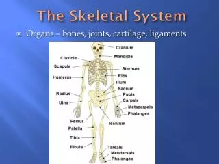
The Skeletal System. Organs – bones, joints, cartilage, ligaments. Functions. A. Support – hard framework that supports and anchors all soft organs of the body – Ex. Legs act as pillars to support trunk, rib cage supports thorax wall

The Skeletal System
E N D
Presentation Transcript
The Skeletal System • Organs – bones, joints, cartilage, ligaments
Functions A. Support – hard framework that supports and anchors all soft organs of the body – Ex. Legs act as pillars to support trunk, rib cage supports thorax wall B. Protection – skull protects brain, ribs protect heart/lungs, zygomatic arch protect the eye C. Movement – skeletal muscles, attached to bone by tendons, used the bones as levers to move the body and it’s parts; arrangement of bones and the design of joints determine the types of movement possible
Functions (con’t) D. Storage • Fat stored in the internal cavities • Bone matrix stores minerals (Ca+2) – “deposits” and “withdrawals” of minerals to and from bones goes on almost continuously E. Blood cell formation (hemopoiesis) • Carried on in red bone marrow; hemopoietic tissue – Fig. 6-5 • Hemopoietic tissue is found in the ends of long bones • Transformed to yellow bone marrow, an inactive fatty tissue, as person ages
Classification of Bones A. Classified according to shape B. Contain different proportions of 1. Compact bone (smooth & homogeneous) 2. Spongy (cancellous) bone – spaces & trabeculae (beams)
Classification of Bones • Long bones 1. Longer than they are wide 2. Consists of a shaft plus two ends 3. Primarily compact bone; but may contain substantial amounts of spongy bone
Classification of Bones D. Short bones 1. Roughly cube-like – ex. Wrist/ankle 2. Mostly spongy bone; compact bone only provides a thin surface
Classification of Bones E. Flat Bone 1. Thin, flattened and usually a bit curved – sternum, ribs, most skull bones 2. Two roughly parallel compact bone surfaces with a layer of spongy bone between
Classification of Bones F. Irregular bones – some skull, hip, vertebrae 1. Fit none of the preceding classes 2. Complex shapes 3. Mainly spongy bone enclosed by thin layers of compact bone G. Sesamoid bones –a special type of short bone embedded within a tendon; ex. - patella
Bone Structure – 2 levels A. Gross Anatomy – What you can see with the naked eye - Fig. 6-1 B. Microscopic Anatomy – Fig. 6-2, 6.3
Gross Anatomy Long Bone – most have the same basic structure 1. diaphysis (shaft) – hollow tube of hard compact bone 2. medullary cavity – hollow area; contains yellow bone marrow 3. epiphyses – bone end or extremities; usually more expanded than diaphysis; thin layer of compact bone forms exterior; interior spongy bone filled with red bone marrow 4. epiphyseal line/plate – remnant of cartilage present at junction of diaphysis & epiphyses in young bones; growth are that allows bones to lengthen 5. articular cartilage – found where long bones articulate (join); cushions the bone ends and absorbs stress during joint movement 6. periosteum – outer surface of diaphysis; richly supplied with nerve fivbers, lymphatic vessels, and blood vessels which enter via nutrient canals 7. endosteum – fibrous membrane that lines medullary cavity
Microscopic Anatomy Compact bone – Fig. 6-2, 6-3 1. Haversian system – structural unit; circular 7 tubelike; composed of calcified matrix arranged in multiple layers (one inside the other – like an onion) 2. osteocytes (bone cells) – regulate the removal of calcium from bone matrix Osteocyte (within lacuna)
Microscopic Anatomy Spongy bone 1. Trabeculae – structural unit 2. Osteocytes – only a few cell layers thick; no Haversian system 3. Nutrients reach osteocytes by diffusion
Microscopic Anatomy Cartilage – Fig. 6-4 1. Fibers embedded in gel (not calcified matrix) 2. Chondrocytes (cartilage cells) 3. Cartilage contains no blood vessels; nutrients diffuse through matrix 4. Function a. supports & reinforces b. cushioning properties c. resists compressive stress (articular cartilage)
Bone Development(osteogensis) – Fig. 6-5 A. Intramembranous ossification – flat bones form from fibrous membrane – ex. Skull, clavicle, ribs B. Endochondral ossification – bone formation from hyaline cartilage structures; most bones form this way; osteoblasts – bone forming cells osteoclasts – bone reabsorbing cells
EndochondralOssifcation – Fig. Cartilage model is the starting point
EndochondralOssifcation – Fig. Formation of a bone collar around the shaft of the hyaline cartilage model
EndochondralOssifcation – Fig. Cartilage matrix calcifies; chondrocytes die
EndochondralOssifcation – Fig. Invasion of internal cavities by periosteal bud and spongy bone formation (3 mo. embryo)
EndochondralOssifcation – Fig. As the primary ossification enlarges, osteoclasts break down spongy bone & form medullary cavity
Ossification of epiphyses - development of secondary ossification centers in epiphyses; cartilage begins to become bone - when complete cartilage remains only at epiphyseal surfaces (articlular cartilage) and at the epiphyseal plate
EndochondralOssifcation – Fig. 6-5 Bone growth continues during infancy & youth - long bones lengthen at epiphyseal plate - long bones thicken by a process called appositional growth (inside breaks down at a slower rate than exterior builds up) - some facial bones (nose, mandible) grow throughout life
The Skeleton – 206 bones Axial skeleton – forms long axis of body & includes the bones of the skull, vertebral column, and rib cage Appendicular skeleton – bones of upper and lower extremeties and girdles (shoulder/hip)
The Axial Skeleton – 80 bones A. Skull – body’s most complex bony structure – Fig. 6-8 1. Cranial bones (8) a. site of attachment of head muscles b. enclose & protect brain & organs of hearing & equilibrium
The Axial Skeleton – 80 bones 2. Facial bones (14) a. form framework of face b. hold eyes in an anterior position c. provide cavities for the organs of taste & smell and openings for the passage of air & food d. secure teeth e. anchor the facial muscles of expression
The Axial Skeleton – 80 bones 3. Middle ear bones (6) – used in sense of hearing
The Axial Skeleton – 80 bones 4. Sutures – interlocking joints of skull bones
The Axial Skeleton – 80 bones B. Vertebral column – 26 irregular bones that form a flexible curved rod that supports the body trunk 1. Provides attachment points for ribs & muscles of back 2. Division of spine – curvature increases strength, resilience & flexibility of spine, making it function like a spring rather than a rod
The Axial Skeleton – 80 bones C. Thorax – 12 pairs of ribs (both male & female), sternum, thoracic vertebrae, costal cartilage 1. Forms protective cage around thoracic organs 2. Supports shoulder girdles & upper limbs 3. Provides attachment points for the muscles of the back, chest, & shoulders 4. Intercostal spaces – occupied by inter- costal muscles which elevate & depress during breathing
The Appendicular Skeleton 126 bones A. Adapted to carry out movement B. Pectoral (shoulder) girdle – clavicle, scapula C. Arm/hand – humerus, radius, ulna, carpals, metacarpals, phalanges D. Pelvic girdle – coxal bones (ilium, ischium, pubic) E. Leg/feet – femur, tibia, fibula, patella, tarsals, metatarsals, phalanges
Male & Female Skeletal Differences A. Most male skeletons are larger (no great functional importance) B. Structural difference in pelvis 1. Male - narrower 2. Female – structured to cradle baby; broader, shallower, lighter, rounder C. Pelvic brim 1. Male – basically heart shaped 2. Female – wider, oval from side to side D. Coccyx 1. Male – narrow, longer; less movable; curves ventrally 2. Female – wider, shorter; more movable; straighter
Articulations (joints) • Two different ways to classify Structural classification – based on material that binds fibrous cartilaginous synovial Functional classification – based on amount of movement
Articulations Synarthroses – immovable joints; fibrous connective tissue grows between the articulating bones; - ex. Sutures of cranial bones
Articulations Amphiarthroses – slightly moveable; cartilage or fibrous tissue connects articulating bones – ex. Symphysis pubis, ligaments, fibrous membrane between radius & ulna
Articulations Diarthroses – allow considerable movement; Fig. 6-20, 6-21, Table 6-7 1. Ball & socket a. shoulder & hip joints b. this type of joint permits the widest range of motion
Articulations • Diarthroses – allow considerable movement; Fig. 6-20, 6-21, Table 6-7 2. Hinge joints a. elbow & knee, fingers, toes b. movement in 2 directions – flexion (bending), extension (straightening)


