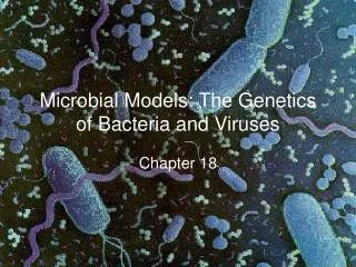
MICROBIAL MODELS
Viruses and Bacteria. MICROBIAL MODELS. Chapter 19 and 27. VIRUSES. No fossil record. When did they arise? . Viral Genomes Capsids and Envelopes Bacterial viruses (Bacteriophages) Animal Viruses Viral Reproduction - Phage Reproduction -Animal Virus Reproduction

MICROBIAL MODELS
E N D
Presentation Transcript
Viruses and Bacteria MICROBIAL MODELS Chapter 19 and 27
VIRUSES No fossil record. When did they arise? • Viral Genomes • Capsids and Envelopes • Bacterial viruses (Bacteriophages) • Animal Viruses • Viral Reproduction - Phage Reproduction -Animal Virus Reproduction • Important Viral Diseases in Animals • Emerging Viruses • Viruses and Cancer • Viroids and Prions
Viral Genomes Depending on the type of virus, it can have either a: • Double-stranded DNA, or • Single-stranded DNA, or • Double-stranded RNA, or • Single-stranded RNA The nucleic acid can be either in a single linear form, or a circular form. Each virus has a “host range” which means it can only infect certain types of hosts.
Capsids And Envelopes • The capsid is a protein shell enclosing the viral genome. Capsids are built of a large number of protein subunits called capsomeres, but with limited diversity. • Some viruses have viral envelopes, membranes cloaking their capsids. These envelopes are derived from the membrane of the host cell. They also have some viral proteins and glycoproteins.
Haemophilus Influenza A HIV Epstein Barr Virus Herpes Simplex Herpes simplex and Epstein Barr belong to a group of enveloped ds DNA viruses called Herpesviridae The Influenza virus belongs to a group of enveloped ss RNA viruses called Orthomyxoviridae HIV belongs to a group of enveloped ss RNA viruses called Retroviridae
Bacteriophages A T-even phage, infecting an E.coli cell
Reproduction of Bacteriophages • Lytic Cycle – virulent viruses - In the lytic cycle the phage reproductive cycle culminates in the death of the host, when the bacterium lyses and releases the phages produced to infect others. • Virulent phages reproduce only by a lytic cycle. • Lysogenic Cycle – temperate viruses - Temperate phages, like phage lambda, use both lytic and lysogenic cycles. During a lytic cycle, the viral genes immediately turn the host cell into a virus-producing factory, and the cell soon lyses and releases its viral products. - During the lysogenic cycle, the viral DNA molecule, is incorporated by genetic recombination into a specific site on the host cell's chromosome. In this prophage stage, one of its genes codes for a protein that represses most other prophage genes. Every time the host divides, it also copies the viral DNA and passes the copies to daughter cells. Occasionally, the viral genome exits the bacterial chromosome and
Lysogenic Cycle A temperate phage that has integrated its genome into that of its host
Animal Viruses • Depending on virus, genomes can be • dsDNA • ssDNA • dsRNA • ssRNA – retroviruses • Refer to Table 18.1 in book
Animal Viruses Very Diverse! They differ in: • The type of nucleic acid they contain • They differ in the presence or absence of a membranous envelope. Viruses equipped with an outer envelope use the envelope to enter the host cell.
The HIV virus is a Retrovirus They also have an enzyme associated with the RNA called Reverse Transcriptase, which transcribes a DNA molecule off the RNA template and then converts it into a double-stranded DNA. This can then be incorporated into the host cell’s genome. Retroviruses have 2 identical single-stranded RNA molecules for their genetic material.
The Ebola Filovirus The AIDS / HIV VIRUS Emerging Viruses mutationRNA viruses have high mutation rates because replication of their nucleic acid lacks proofreading. Influenza strainsspread of existing viruses from one species to anotherIt is estimated that about three-quarters of new human diseases have originated in other animals.For example, hantavirus, which killed dozens of people in 1993, normally infects rodents, especially deer mice. Dissemination of a viral disease from a small, isolated population.AIDS, present only in small populations in Africa, went unnamed and unnoticed for decades before spreading around the world. Affordable international travel, blood transfusion technology, sexual promiscuity, and the abuse of intravenous drugs, allowed a previously rare disease to become a global scourge. Enveloped ss RNA virus
The bird influenza virus is very closely related to the human Influenza-A virus, an enveloped ss RNA virus More Emerging Viruses The SARS virus belongs to a genus of enveloped ss RNA viruses called Coronaviridae. This virus is also called SARS-CoV
Viroids and Prions Viroids, smaller and simpler than even viruses, consist of tiny molecules of naked circular RNA that infect plants. Their several hundred nucleotides do not encode for proteins but can be replicated by the host's cellular enzymes. Prions are infectious proteins that spread a disease. They are thought to cause several degenerative brain diseases including scrapie in sheep, "mad cow disease," and Creutzfeldt-Jacob disease in humans. According to the leading hypothesis, a prion is a misfolded form of a normal brain protein, which then converts normal proteins into the prion version.
Viruses living or nonliving? • An isolated virus is biologically inert and yet it has a genetic program written in the universal language of life. • Because viruses depend on cells for their own propagation, they evolved after the first cells appeared, probably fragments of cellular nucleic acids that could move from one cell to another. • A viral genome usually has more in common with the genome of its host than with those of viruses infecting other hosts. • The evolution of capsid genes may have facilitated the infection of undamaged cells. • Candidates for the original sources of viral genomes include plasmids, small, circular DNA molecules that are separate from chromosomes, and transposons, DNA segments that can move from one location to another within a cell's genome. Both are mobile genetic elements.
Proto-Oncogenes and Oncogenes • All cells contain genes called Proto-Oncogenes. • These genes are actually normal genes that give rise to proteins necessary for normal growth of the cell • If however the expression of these proto-oncogenes is up-regulated, their protein products will cause the cell to grow and divide uncontrollably – leading to tumors. • These over expressed proto-oncogenes are now called oncogenes (cancer causing genes)
How do Viruses cause Cancer? • Many viruses incorporate their DNA into the host’s genome along with their own powerful promoters • This causes host RNA polymerase to transcribe the viral DNA at a rapid rate – allowing the virus to make millions of copies of itself. • Sometimes, the viral DNA may get incorporated next to a proto-oncogene • This will cause the proto-oncogene to be over-expressed along with viral genes – leading to tumors • Some retroviruses even carry oncogenes (picked up from previous hosts) in their genome and these get incorporated into the host genome along with other viral genes
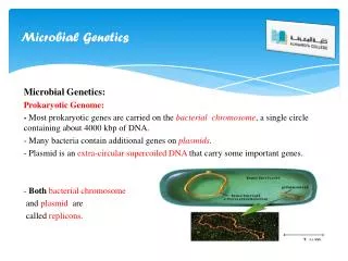
The Genetics of Bacteria • Prokaryotes • Reproduction is called Binary Fission • Single circular DNA • Plasmids and Episomes • Genetic Recombination - Transformation - Transduction - Conjugation • Transposons • Simple and Composite insertion sequences • Operons - lac operon - trp operon
The New System • Adds a level above Kingdom called Domain • There are three domains – two of which contain prokaryotes and one contains eukaryotes
PROKARYOTES No nucleus or membrane-bound organelles. Domain Eubacteria(Bacteria) Domian Archaebacteria
Prokaryotic Flagella Prokaryotic flagella are composed of chains of the protein flagellin, are attached to another protein that forms a hook that is inserted into the basal apparatus. The flagellum spins around this basal apparatus in a circular motion, which is quite different from the motion of the eukaryotic flagella.Remember what eukaryotic flagella are made of?
Prokaryotes are grouped into 4 categories based on how they obtain their energy and carbon • Photoautotrophs– harness light to synthesize organic compounds from carbon dioxide – cyanobacteria (Also plants and algae – eukaryotes) • Chemoautotrophs – use CO2 to synthesize organic compounds, but instead of harnessing light energy, they obtain energy by oxidizing inorganic substances such as H2S and NH3 (nitrogen metabolism) • Photoheterotrophs – Harness light energy, but have to derive their carbon from organic compounds instead of from CO2 • Chemoheterotrophs – Consume organic molecules for both energy and carbon – most common mode of nutrition in prokaryotes and eukaryotes such as protists, fungi, animals and even certain plants!
Chemoheterotrophs • Saprobes – decomposers (get nutrition from dead organic matter) • Parasites – absorb nutrients from living hosts
Chemoautotrophs • Nitogen fixing bacteria – N2 to ammonia and ammonium • Nitrifying bacteria – NH3 to NO2(nitrite) and NO3(nitrate) • Denitrifying bacteria – NO3(nitrate) to N2 gas
Metabolism and Oxygen • Obligate aerobes – absolutely must have O2 • Facultative anaerobes – aerobic if O2 is present, fermentation if it is not • Obligate anaerobes –hate O2 and die if exposed to it (“Airing-out kills germs”) • Aerobic respiration uses O2 as the final electron acceptor • Anaerobic respiration uses a different inorganic molecule like sulfur or iron
Chromatium, a proteobacterium, and the green sulfur bacterium, Chlorobium. These photoautotrophs do not evolve oxygen during photosynthesis, but rather produce sulfur as a biproduct of photosynthesis. Rhizobium bacteria Eubacteria – Modern Bacteria • Proteobacteria – the most diverse group of bacteria • Chemoautotrophic proteobacteria -Nitrogen-fixing bacteria – live in soil, helpful to plants (Rhizobium) • Photoautotrophs and photoheterotrophs (Purple bacteria) – with chlorophyll in their plasma membranes – use H2S instead of H2O for their version of photosynthesis (Photolysis) and release sulfur instead of oxygen • Chemoheterotrophic proteobacteria – enteric bacteria such as E.coli, Vibrio cholerae, Salmonella. Mostly rod-shaped (also classified as Gram negative, facultative anaerobes)
Cyanobacteria Streptococcus pneumoniae Spirochetes Eubacteria – Modern Bacteria, cont’d 2. Cyanobacteria – photosynthesizers, contain chlorophyll 3. Gram positive bacteria – Clostridium, Bacillus (both spore-producing), Mycoplasma pneumoniae, streptococcus 4. Gram negative bacteria – common disease causing bacteria like, E.coli, Salmonella, staphylococcus, etc. 5. Spirochetes – Usually pathogenic. Treponema pallidum causes syphilis, Borrelia burgdorferi causes Lyme disease. • Chlamydias – animal parasites. Chlamydia trachomatis source of STD and common cause of blindness in the world. • Mycoplasma – the smallest free-living cells, cause walking pneumonia in humans
Gram-staining • Gram-negative bacteria are those that do not retain crystal violet dye in the Gram staining protocol. Gram-positive bacteria will retain the dark blue dye after an alcohol wash, whereas Gram-negative bacteria do not. In a Gram stain test, a counterstain (usually safranin) is added after the crystal violet, which colors all Gram-negative bacteria a red or pink color. The test itself is useful in classifying two distinctly different types of bacteria based on structural differences in their cell walls. • Many species of Gram-negative bacteria are pathogenic, This pathogenic capability is usually associated with certain components of Gram-negative cell walls, in particular the lipopolysaccharide (also known as LPS or endotoxin) layer.
Gram stained cerebrospinal fluid showing gram-positive anthrax baccilli (purple rods) – these are gram + (blue) Gram- Enteric E.coli bacilli stained with Safranin counter stain (pink) Gram Staining, cont’d.
Most Common Eubacteria Shapes • Spherical (cocci) • Rod-shaped (bacilli) • Spiral
SEM of Streptococcus pyogenes , causes pharyngitis, food poisoning, scarlet fever, rheumatic fever, skin and wound infections. Group A, Gram + SEM of Neisseria gonorrhoeae, a Diplococcus Gram + Tetrad arrangement Sarcina form cubes of eight (2 tetrads) Cocci Arrangements SEM of Staphylococcus aureus - causes food poisoning, toxic shock syndrome and skin and wound infections such as scalded skin syndrome, scarlet fever. Gram +
Bacilli are rod-shaped bacteria. Bacilli all divide in one plane producing a bacillus, streptobacillus, or coccobacillus arrangement. Bacilli are aerobes and produce spores that allow them to lie dormant for long periods of time (anthrax) Bacilli Arrangement
Spirillum - rat-bite fever SEM of Vibrio cholerae, facultatively anaerobic, curved (comma-shaped), rod prokaryote; causes Asiatic cholera. Gram - SEM of the spirochete Leptospira interrogans causes many diseases including meningitis Gram - spirochete Borrelia burgdorferi (arrows) in a blood smear causes Lyme disease. Gram - Spirals
Replication of Bacterial Chromosome The major component of the bacterial genome is one double-stranded, circular DNA molecule. Tight coiling of the DNA results in a dense region of DNA, called the nucleoid, not bound by a membrane.Many bacteria have plasmids, much smaller circles of DNA.Bacterial cells divide by binary fission.
Plasmids • Plasmids, including the F plasmid and R plasmids are small, circular, self-replicating DNA molecules. The F Plasmid is responsible for giving a bacterium its “maleness” and allows it to develop a sex pilus. Episomes, are a genetic element that can exist as a plasmid or as part of a chromosome. Episomes, like the F plasmid, can undergo reversible incorporation into the cell's chromosome. Temperate virus genomes also qualify as episomes. Plasmid genes are advantageous in stressful conditions. Recombination exchanges segments of DNA. This recombinant bacteria has genes from two different cells. Antibiotic resistance. • The genes conferring resistance are carried by plasmids, specifically the R plasmid (R for resistance). • Some of these genes code for enzymes that specifically destroy certain antibiotics, like tetracycline or ampicillin. • When a bacterial population is exposed to an antibiotic, individuals with the R plasmid will survive and increase in the overall population. • Because R plasmids also have genes that encode for sex pili, they can be transferred from one cell to another by conjugation.
Transformation • Transformation is the alteration of a bacterial cell's genotype by the uptake of naked, foreign DNA from the surrounding environment. For example, harmless Streptococcus pneumoniae bacteria can be transformed to pneumonia-causing cells, when a live nonpathogenic cell takes up a piece of DNA that happens to include the allele for pathogenicity from dead, broken-open pathogenic cells.
Transduction • A phage infects a susceptible bacterium and injects its DNA into the host. • The phage DNA proceeds to TAKE OVER the host's entire metabolism, turning it into a PHAGE-FACTORY. • It then directs the synthesis of PHAGE PARTS or COMPONENTS, including phage DNA. • As a final step in the phage life-cycle, the VARIOUS COMPONENT PARTS in the cytoplasm are ASSEMBLED into COMPLETE PHAGE and the cell is broken open or LYSED, releasing the newly made phage particles. • During the assembly process, occasional RARE MISTAKES are made and bits of the host's bacterial DNA become incorporated INTO THE NEW PHAGE. • Since there is NO ROOM FOR PHAGE DNA in these situations DEFECTIVE PHAGE are made. • However, these defective phage still have the ability to subsequently bind and INSERT their DNA into a host bacterial cell. But, since these defective phage contain only bacterial DNA, an infection DOES NOT OCCUR. • Recombination can take place between the previous host's DNA and the new host's DNA and any alleles carried into the host by the defective phage can become INCORPORATED into the host's DNA, thereby CHANGING ITS GENOTYPE
Conjugation Conjugation transfers genetic material between two bacterial cells that are temporarily joined. One cell ("male") donates DNA and its "mate" ("female") receives the genes. A sex pilus from the male initially joins the two cells and creates a cytoplasmic bridge between cells. "Maleness," the ability to form a sex pilus and donate DNA it is a result of an F factor as a section of the bacterial chromosome or as a plasmid.
Archaebacteria (cont’d) Extreme Halophiles Love salty environments Methanogens Hate O2, make CH4 Thermoacidophiles Or Extreme thermophiles Love heat and / or acidity