DMSO
Ciglitazone. DMSO. 0 0.5 1 1.5 0 0.5 1 1.5 h. IRF-1. GAPDH. 1.2. DMSO. Ciglitazone. 1. 0.8. Relative IRF-1 mRNA Level. 0.6. 0.4. 0.2. 0. 0. 0.5. 1 hr. Time post actinomycin D treatment.

DMSO
E N D
Presentation Transcript
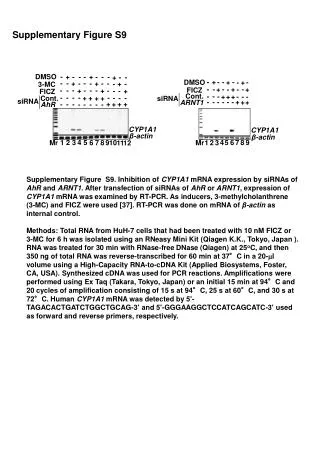
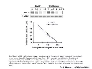
Ciglitazone DMSO 0 0.5 1 1.5 0 0.5 1 1.5 h IRF-1 GAPDH 1.2 DMSO Ciglitazone 1 0.8 Relative IRF-1 mRNA Level 0.6 0.4 0.2 0 0 0.5 1 hr Time post actinomycin D treatment Fig. I. Decay of IRF-1 mRNA in the presence of actinomysin D. Human aortic smooth muscle cells were incubated with or without 10mmol/L of ciglitazone for 2 h, and de novo IRF-1 transcripts were inhibited by the addition of actinomysin D (5mg/ml). Total RNA was isolated at 0, 0.5, 1, and 1.5h after administration of actinomysin D. A representative Northern-blotting analysis (top) and the quantitative graph (bottom) of three experiments are shown. The relative values were normalized by GAPDH. Values for IRF-1 mRNA are expressed as means SD (n=3) in the bottom panel. Fig. I. Lin et al. ATVB/2003/003848
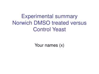
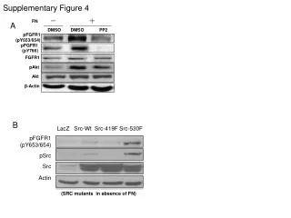
4.5 4 p<0.01 3.5 3 2.5 Relative Luciferase Activity 2 1.5 1 0.5 0 Ciglitazone PcDNA3.1 PPAR 0 5 0 5(mol/L) Figure II. PPAR Activation Increased the transactivation of IRF-1 Promoter. CV-1 cells were co-transfected with pIRF-1-2kbLuc, pCMV-GFP, and pcDNA3.1-PPAR expression plasmid or pcDNA3.1 vector. GFP was used as the control for the transfection efficiency. The luciferease activities normalized by GFP activity were expressed as meansSD (n=4). Fig. II. Lin et al. ATVB/2003/003848


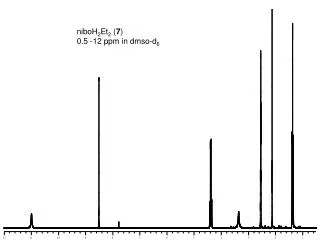
![1 H NMR (500 MHz, d 6 -DMSO) [Cu(Thr)(Byp)Cl]·H 2 O (1)](https://cdn1.slideserve.com/3407140/slide1-dt.jpg)