Download
1 / 18
180 likes | 365 Views
Proteins. proteins are the most active macromolecules in living cells. they are polymers of basic building blocks called amino acids typical protein contains 200-300 amino acids largest protein „titin“ = 27 000 aa). Functions of Proteins.

E N D
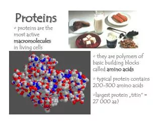
Proteins • proteins are the most active macromoleculesin living cells • they are polymers of basic building blocks called amino acids • typical protein contains 200-300 amino acids • largest protein „titin“ = 27 000 aa)
Functions of Proteins • Structural – serve as support molecules within and between cells – e.g. collagen in skin; muscle protein; cell membranes; • Enzymes– all enzymes are proteins; catalyse almost all biochemical reactions • Transport– carry molecules such as lipids (lipoproteins HDL/LDL) and O2 (haemoglobin) • Antibodies– immune system‘s main defence molecule; attach to toxins, viruses, bacteria, etc.
Amino Acids • about 20 common amino acids • link together side-by-side amino group to carboxyl group • linkage is called peptide bond The R-group is what gives an amino acid its identity, its properties. Each aa is unique though there are some fundamental types of aa. Types of AA:acidic, basic, hydrophobic, water soluble, aromatic, etc
Four Levels of Protein Structure • As amino acids link together, a polypeptide chain is formed • As polypeptide chain grows, it twists and turns and forms various patterns, eventually forming a complex 3-D structure The primary structure of a protein is the sequence of amino acids that make up that protein – their names, positions, and the exact locations for disulfide bridges.
The primary structureof the protein hexokinase (from the yeast speciesSaccharomyces cerevisiae) amino end A A S X D X S L V E V H X X V F I V P P X I L Q A V V S I AT T R X D D X D S A A A S I P M V P G W V L K Q V X G S Q AG S F L A I V M G G G D L E V I L I X L A G Y Q E S S I X AS R S L A A S M X T T A I P S D L W G N X A X S N A A F S S X E F S S X A G S V P L G F T F X E A G A K E X V I K G Q I T X Q A X A F S L A X L X K L I S A M X N A X F P A G D X X X X V A D I X D S H G I L X X V N Y T D A X I K M G I I F G S G V N A A Y W C D S T X I A D A A D A G X X G G A G X M X V C C X Q D S F R K A F P S L P Q I X Y X X T L N X X S P X A X K T F E K N S X A K N X G Q S L R D V L M X Y K X X G QX H X X X A X D F X A A N V E N S S Y P A K I Q K L P H F D L R X X X D L F X G D Q G I A X K T X M K X V V R R X L F L I A A Y A F R L V V C X I X A I C Q K K G Y S S G H I A A X G S X R D Y S G F S X N S A T X N X N I Y G W P Q S A X X S K P I X I T P A I D G E G A A X X V I X S I A S S Q X X X AX X S A X X A carboxyl end 457 amino acids
The secondary structure of a protein is/are the pattern(s) that form along the polypeptide chain due to H-bonding between the R-groups on the amino acids. • There are two main patterns – the helical coil (helix) and the pleated sheet;
The tertiary structure of a protein is its 3-dimensional shape. • Bonding forces of many types (attractive and repulsive) between the amino acids cause the mature polypeptide chain to assume a final shape. • The shape of a protein is directly related to its function – if a protein loses its shape, it cannot carry out its function. • Enzymes are controlled by slightly altering their shape, often only in one „sensitive“ area of the whole protein. • Large-scale disruption to a protein‘s shape is called denaturation. The amino acids of denatured proteins may still be attached together, but the protein cannot function.
Typical Ways of Viewing Proteins Ribbon Model Space-filling Model
Many proteins show an even more complex level of structure, where two or more polypeptide chains join together to form a functional protein. This fourth level of structure is called a quaternary structure. Two different Views of Haemoglobin


