Download
1 / 30
330 likes | 491 Views
Learn about the various cardiac defects in children, the classification, hemodynamic features, clinical manifestations, and recommended interventions like surgery or catheterization. Understand the importance of prompt intervention for conditions like ASD, VSD, and Tetralogy of Fallot.

E N D
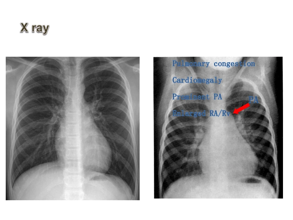
PA X ray Pulmonary congestion Cardiomegaly Prominent PA Enlarged RA/Rv
ECG Right axis deviation RV hypertrophy
心电图 Incomplete Right bundle branch block: V1rsR` QRS < 0.12sec
Catheterization pressure RV PA Oxygen saturation CV 1 RA2 RV3 PA4 1 4 2 3
Angiography Anomalous pathway interatrial
Treatments Surgery: 4~5y/r Intervention is recommended as the first choice in many cases: needs: the site of defect the margin of the defect
Introduction AtrialSeptal Defects Ventricular Septal Defects Patent DuctusArteriosus Tetralogyof Fallot Pulmonary Stenosis
Phathological classification The ventricular septum is divided into : a small membranous portion a large muscular portion the inlet septum the trabecular septum the outlet septum
Phathological classification Membranous defect: most common 70%
Phathological classification Infundibular defect: About 5~7%
Phathological classification Muscular defect: About 13~15%
Size classification Nonrestrictive VSD
Hemodynamic Features : CV RA RV PA LA LV Ao Pulmonary Circulation
Hemodynamic Features : CV RA RV PA LA LV Ao Pulmonary Circulation
Clinical Manfestations small:asymptomatic,Roger disease moderate to large: delayed growth and development decreased exercise tolerance repeated pneumonia CHF
Clinical Manfestations Automatic closure occurs in about 20% cases within 2 y/r A few cases have aortic insufficiency Large VSDs suffer from Eisenmenger Syndrome in earlier stage Infectious endocarditis: a main complication
Physical Examination Increased P2 intensity Grade 3-6 SM is audible at 3-4 left sternal border Systolic thrill Enlarged cardiac border 心尖区闻及DM
X ray • Pulmonary congestion • cardiomegaly • Prominent pulmonary marking • Enlarged LV RV • Enlarged LA
ECG small: LV hypertrophy large: ventricular hypertrophy Depressed ST-T
Catheterization pressure RV PA Oxygen saturation CV 1 RA2 RV3 PA4 1 4 2 3