Download
1 / 63
630 likes | 950 Views
DON’T FAIL MY HEART. Physiology, Pathology and Treatment of Heart Failure. Objectives. To understand how a normal heart develops heart failure, learning the physiologic compensatory mechanisms that play roles in preventing and/or delaying progression to a failing heart.

E N D
DON’T FAIL MY HEART Physiology, Pathology and Treatment of Heart Failure
Objectives • To understand how a normal heart develops heart failure, learning the physiologic compensatory mechanisms that play roles in preventing and/or delaying progression to a failing heart. • To learn the different symptoms and signs that signal heart failure and understanding how these develop in the course of the disease; and • To acquire knowledge on how to approach treatment of heart failure addressing the different pathologic insults that lead to the development and progression of the failing heart.
Definition Heart Failure is a clinical syndrome that occurs in patient who, because of an INHERITED or acquired abnormality of cardiac structure and/or function, develop a constellation of clinical symptoms (dyspnea and fatigue) and signs (edema and rales) that lead to frequent hospitalizations, a poor quality of life, and a shortened life expectancy.
Burden of the disease • Prevalence in the adult population in developed countries is 2%. • Prevalence rises with age and affects 6-10% of people over the age of 65. • Categorized into 2 groups: • HF with a depressed EF (systolic failure) • HF with preserved EF (diastolic failure)
Take – off case • ZA • 75 years old • Female • Widow • Roman Catholic • From Tondo • Known hypertensive for 50 years • Known Diabetic for 15 years
History of Present Illness 9 days PTA • Started to complain DOB on excertion described as drowning. • Difficulty climbing 1 flight of stairs • 2-3 pillow orthopnea • Intermittent chest heaviness radiating to the upper back. • Decreased urine output from almost 5 glasses/day to 2 cups/day. • Bipedal edema non-pitting noted • Facial edema noted • Decreased appetite
Day of Admission • Bipedal edema, pitting persisted • Facial edema also persisted • 2-3 pillow orthopnea still noted • Intermittent chest heaviness radiating to the upper back • Decreased urine output still approx. 1-2 cups per day. • Bloatedness which resulted to decreased appetite • Prompted consult to a DM physician in this institution • Noticed by the physician to have abdominal enlargement • Patient was then advised for admission
Past Medical History • 1958 - HTN (UBP= 120-130/90; Highest BP= 160) • Combizar 100mg/25mg/tab, 1 tab OD • Atenolol 100mg/tab, 1 tab OD • Clopidogrel 75mg/tab, 1 tab OD • 1997 – DM type II • HUM 70/30 = 28 ‘u’ AM; 14 ‘u’ PM • 1990 – S/p TAB for myoma Uteri
1995 – S/P cholecystectomy for cholecystitis • 2004 – Mass excision on popliteal area • 2006 – Bronchitis • Hyperurecemia – allopurinol 100mg/tab, 1 tab OD • Dyslipidemia – Simvastatin 40mg/tab, 1 tab qHS
Family History • (+) HTN – mother • (+) Kidney disease – sister • (+) Stroke – sister • (+) TB – mother • (+) DM – mother
Physical Exam on Admission • VS: • Anictericsclerae, pink palpebral conjunctivae, (+) Prominent neck veins, (-) CLADS, (-) TPC • ECE, (+) bibasal crackles, (-) wheezing • AP, distinct S1 and S2, NRRR, (-) murmur • Globular, (+) fluid wave, (-) caput medusae, (-) Bruits • FEP, (+) bipedal edema, pitting
ECG • NSR • Freq. PVCs in singles • Left atrial abnormality • HBa1c: 7.5% • CBG: 52
Chest X-ray – mild bilateral pulmonary congestion – biventricular cardiomegaly – atherosclerotic aorta • 2D Echom
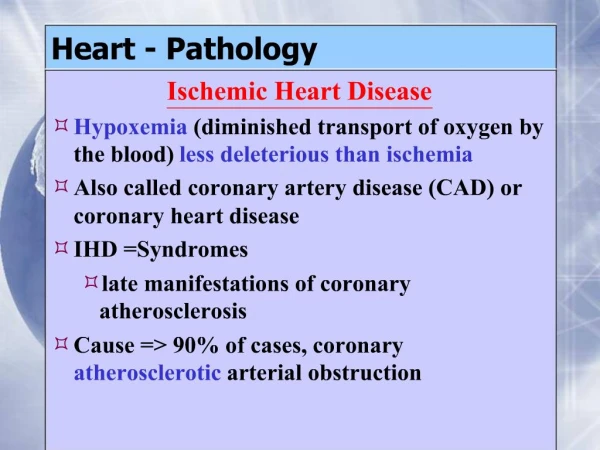
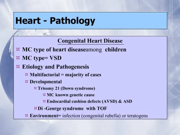
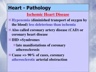
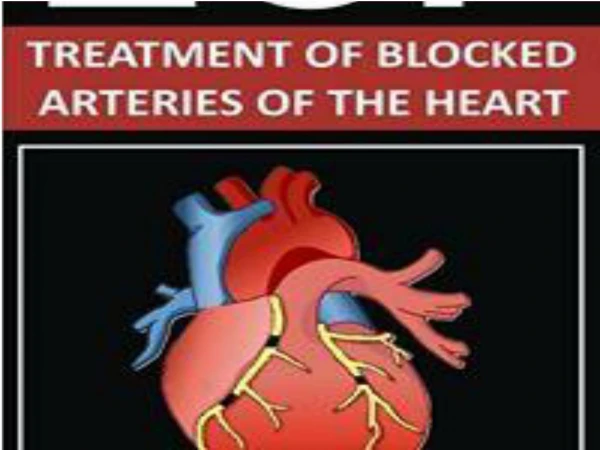
Underlying Cause • Ischemic Heart Disease • Cardiomyopathies • Congenital, Valvular Hypertensive Heart Disease
Precipitating Cause • Infection • Arrhytmia • Physical, Dietary, Fluid, Environment • Myocardial Infarction • Pulmonary Embolism • Anemia • Thyrotoxicosis and Pregnancy • Aggravation of Hypertension • Rheumatic, Viral and other forms of Myocarditis • Infective Endocarditis
Heart Failure • Systolic Dysfunction – Depressed EF • Diastolic Dysfunction – EF Preserved
Hemodynamic Derangement in HF • Reduction in Cardiac Reserve • Increased ventricular diastolic pressure
Systolic Dysfunction • Main Pathology: Decreased Cardiac Output
4 Major Determinants of the Systolic Function of the Heart and the Cardiac Output • Contractile State of the Myocardium • Preload • Afterload • Heart Rate
Diastolic Dysfunction • Main Pathology: Impaired Ventricular Filling
Contractility Preload Afterload Myocardial Fiber Shortening LV Size Heart Rate Stroke Volume Cardiac Output TPR Arterial Pressure
Compensatory Mechanisms to a Decreased Cardiac Output • Increased Sympathetic Activity • Increased Heart Rate • Increased Myocardial Contractility • Increased Venous Tone
Compensatory Mechanisms to a Decreased Cardiac Output • Activation of the RAA System
Compensatory Mechanisms to a Decreased Cardiac Output • Secretion of AVP