Download
1 / 44
450 likes | 469 Views
Oocyte quality and It’s influence on Embryo Cryopreservation Survival and Outcome. Başak Balaban, BSc VKV American Hospital Assisted Reproduction Unit, Istanbul, Turkey. AMER IC AN HOSPITAL. OOGENESIS.

E N D
Oocyte quality and It’s influence on Embryo Cryopreservation Survival and Outcome Başak Balaban, BSc VKV American Hospital Assisted Reproduction Unit, Istanbul, Turkey AMERICAN HOSPITAL
OOGENESIS • The ability of an embryo to implant, sustain and give rise to a viable offspring is unquestionably rooted to oogenesis • During oogenesis significant variations are known to exist among oocytes **Meiotic competence (ability to undergo meiotic maturation) **Developmental competence (support later embryonic development
GOOD QUALITY OOCYTE? • Mature oocyte that gives rise to the best quality embryo with the highest implantation potential • Viable, genetically normal oocyte that gives rise to an healthy embryo and healthy offspring
FACTORS AFFECTING OOCYTE QUALITY • Intrinsic factors **- Chromosomal abnormalities, genetic defects,suboptimal characteristics of the oocyte cytoplasm • Extrinsic factors **- Stimulation protocols - Culture conditions (Ebner et al.2005, protocols and factors spesific to the laboratory - Impact of nutrition??(Moderately fat animals on high level of feeding were found to be hyperinsulinaemic,and ruminants with this condition are associated with impaired oocyte quality, Adamiak et.al.2005)
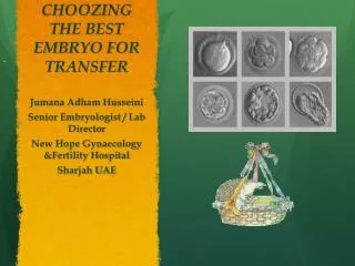
GOOD QUALITY OOCYTEMORPHOLOGY • Perfectly spherical shape • Small Perivitelline Space(PVS) • Regular Zona Pellucida (ZP) • Intact 1st. Polar Body (PB) • Translucent, homogeneously coloured cytoplasm without inclusions Veeck et al. Ann.N.Y.Acad.Sci, 1988 **Only 34% of all oocytes retrieved are of ideal morphology
Markers of oocyte quality /viabilityin relation to morphology • Cumulus-corona morphology • 1st.Polar body morphology • Metaphase II (MII) OOCYTE MORPHOLOGY
Cumulus-corona morphology A B C D Ng, Fertil Steril, 1999
Pro: Ng, Fertil Steril, 1999 (IVF) Lin et al.,JARG 2003 (IVF) Con: Rattanachaiyanont, Fertil Steril, 1999 (ICSI) Hammitt, JARG, 1992 (IVF) Hammitt, Fertil Steril, 1993 (IVF) Cumulus-corona morphology Highly dependant on embryologist’s experience Asynchrony in 28%
Polar body morphology Oocyte grading according to first polar body morphology Ebner, Fertil Steril, 1999 Ebner, Hum Reprod, 2000 Ebner, Hum Reprod, 2002
Predictive value of 1st.PB morphology evaluation?? • Formation of the 1st.PB is a dynamic procedure and morphological characteristics (fragmentation) of the PBI of human oocytes can significantly change before ICSI and after 16 h in culture. Therefore cannot be used as a diagnostic criteria determining the quality of the subsequent embryo and it’s implantation potential (Verlinsky et al., RBM Online 2003) • Predictive value of 1st.PB morphology is limited,and could not be used as a prognostic factor of embryo quality& PR & IR (Ciotti et al., RBM Online 2004)
MORPHOLOGICAL ABNORMALITIES OF MII OOCYTE • Extracytoplasmic abnormalities **Shape abnormalities (irregular shape of MII oocyte) **ZP abnormalities (dark or thick ZP) **PVS abnormalities (large PVS, PVS granulation) • Cytoplasmic abnormalities **Dark cytoplasmic colour (slight granulation) ** Excessive whole or centrally located granulation **Refractile bodies,sERC or vacuoles in the ooplasm
Effect of extracytoplasmic abnormalities on embryo development&CPR&IR **CON: • De Sutter et al.,Hum.Reprod. 1996 • Balaban et al.,Hum.Reprod. 1998 • H.Hassan-Ali et al.,Hum.Reprod.1998 • Loutradis et al.,F&S 1999 • Ebner et al.,JARG 2001 • Plachot et al.,Gyn. Obst. Fertil 2002 **PRO: • Xia et al.,Hum.Reprod. 1997(CPR not mentioned)
Effect of cytoplasmic abnormalities on embryo development&CPR&IR **PRO • Serhal et al., Hum.Reprod. 1997 • Xia et al.,Hum.Reprod. 1997 • Loutradis et al.,F&S 1999 • Ebner et al.,HumReprod. 2003 • Otsuki et al.,Hum.Reprod. 2004 • Ebner et al.,Hum.Reprod. 2005 **CON • De Sutter et al.,Hum.Reprod. 1996 • Balaban et al.,Hum.Reprod. 1998(severe defects excluded)
Smooth Endoplasmic Reticulum Clusters (sERC) and vacuolization appearance in MII oocytes Otsuki et al.Hum.Reprod.,2004
The presence of sERC is associated with lower chances of successful pregnancy Otsuki et al.Hum.Reprod.,2004
Very low OPR% are obtained with the transfer of embryos obtained from CLCG oocytes Kahraman&Yakin et al.Hum.Reprod.,2000 CLCG-Centrally located granular cytoplasm Fert%+embryo quality: no sig.
Correlation of oocyte morphology with aneuploidy rates Yakin&Balaban et al.,F&S 2007
Effect of cytoplasmic vacuoles throughout embryo development Mean diameter of vaculoes with fertilization :9.8m Mean diameter of vaculoes with FF :17.6 m (p<.05) Cut off value :14 m Patients variables similar between vacuol (+) & (-) cycles Ebner et al. F&S, 2005
Markers of Oocyte Quality/Viability • Examination of the structure of miotic spindle, and zona pellucida of the oocyte by using polirized microscopy
Spindle imaging of the MII oocyte De Santis et al., RBM Online 2005 At the time of ovulation mammalian oocyte is arrested at MII of the meiotic Cell cycle, when chromosomes are tethered by microtubule fibres of the meiotic Spindle. During meiosis and fertilization meiotic spindles are responsible for proper segregation of the nuclear material, and abnormalities in this fragile structure can lead to İnfertility, miscarriage and genetic diseases, such as Down syndrome
Oocyte zona birefringence intensity High zona birefringence Low zona birefringence Montag et al.,RBM Online 2008 Polarization microscopy allows the distinction of three layers within the ZP Inner layer exhibits the highest birefringence(Pelletier 2004). Zona birefringence intensity is higher in conception cycles(Shen 2005)
Abnormalities of oocyte morphology do effect embryo quality and viability in certain cases • Whether morphological abnormalities of the oocyte influence cryosurvival and further development of derived embryos in not known
Aim of the study • To compare survival, blastocyst formation and hatching rates of frozen-thawed day 3 cleavage stage embryos derived from morphologically abnormal or normal metaphase II (MII) oocytes
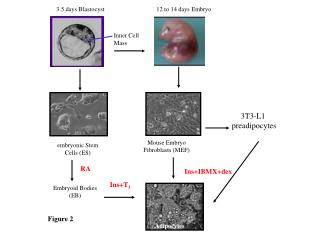
Material and Methods • 5292 day 3 excess embryos of good quality from 964 patients (mean age: 32.4) were frozen and subsequently cultured up to the blastocyst stage after thawing • Fertilization was affected by ICSI (cycles with testicular/epididymal sperm were excluded) • GIII series (Vitrolife-Sweden) for fresh embryo culture, and Freeze/Thaw Kıt-1(1-2 propandiol based- Vitrolife)/with a slow freezing program was used for freezing
Blastocyst development of cryo-thawed top quality day 3 embryos (Single morphological abnormality) Balaban et al.HR 2008 * Sig. diff. from normal MII
Blastocyst development of cryo-thawed good quality day 3 embryos (Single morphological abnormality) Balaban et al.,HR 2008 *Sig.diff. from normal MII
Blastocyst development of cryo-thawed 8 cell embryos (Single morphological abnormality) *Sig.diff. from normal MII Balaban et al.,HR 2008
Blastocyst development of cryo-thawed top quality day 3 embryos (Multiple morphological abnormality) Balaban et al.,HR 2008 *Sig.diff. from normal MII
Blastocyst development of cryo-thawed good quality day 3 embryos (Multiple morphological abnormality) Balaban et al.,HR 2008 *Sig.diff. from normal MII
Blastocyst development of cryo-thawed 8 cell embryos (Multiple morphological abnormality) Balaban et al.,HR 2008 *Sig.diff. from normal MII
Conclusions- I • Presence of extracytoplasmic abnormalities alone does not affect blastocyst development despite decreasing cryosurvival • However, embryos derived from oocytes with vacuolar cytoplasm or central granulation do not seem to bear the potential to develop into good quality blastocyst or to reach hatching stage after cryopreservation • These cytoplasmic abnormalities may be reflections of genetic, epigenetic or metabolic defects in the oocyte
Conclusions-II Embryos with severe cytoplasmic abnormalities comprise around 5% of all embryos suitable for cryopreservation. The formation of a good quality blastocyst, or a blastocyst with the capibility of completing the hatching procedure after thawing of such embryos is ≤1% Although transfer of such embryos may not affect overall success rate of a cryopreservation program, priority should be given to the transfer of embryos derived from oocytes without cytoplasmic abnormalities whenever possible
Conclusions-III • Although it’s difficult to estimate the impact of oocyte morphology on the implantation potential of the derived embryo when multiple embryos are transferred, and when considering the fact that the transition from the gamete to the embryo is a continuum with the difficulty of evaluating the contribution of the oocyte on implantion potential of the derived embryo, it’s shown that the developmental behaviour of frozen-thawed embryos derived from dysmorphic oocytes are similar when compared to results from the literature reported for fresh embryos (Balaban&Urman 2006, Ebner et al., 2006)
Fertilization&embryo quality outcome in relation to the presence/absence of visible spindle in M2 oocytes NA:not applicable, a: p<0.05 Borini et al RBM Online 2005
Why are the results contradictory?? • The dynamics of spindle formation during oocyte maturation were not considered: *Spindle visualization might change within the maturation stages: MI-1st.PB extrution- telaphase I (MS dissepear for 40-60 minutes!!)- MII. Ideal time at least 38 hours after the HCG(Cohen 2004) *The microtubules of the MS are highly sensitive to chemical (hyaluronidase), and physical changes ( temperature& pH variations) that may occur during oocyte handling. Shift of the PB1 position may also be related to physical displacement during denudation • A precise classification spindle imaging should be performed repeatedly: after hyaluronidase treatment and immediately prior to ICSI Rienzi et al.,RBM Onilne 2005, Montag et al.,RBM Online 2006 Not only the presence of the spindle but also the angle to the 1st.PB, retardance and the length can be evaluated to determine the quality of the OOCYTE Rienzi 2003, Rama Raju 2007
M.Spindle visualization and ICSI outcome: Meta-Analysis Petersen C et al.,RBM Online 2009 PN morphology p:0.003, Cleavage: p<0.0001, Blastocysts:p<0.0001
Transfer of the embryos derived from oocytes with HZB reveals higher IR&OPR Montag et al.,RBM Online 2008 Rama Raju( 2007) had also shown sig. higher blastocyst formation from oocytes with higher zona inner layer retardance measurements
35.6% No.of patients: 69 No.of day 5 BT: 49 No.of day 3 ET: 20 LBR: 34.8% IR:29.1% No.dif in PR for day3&5ET Patients with + PR had sig. fewer oocytes not detected (22.6% vs 35.5.%) Ebner &Balaban F&S 2009 in press
p<0.01 Ebner &Balaban F&S 2009 in press