Download
1 / 74
750 likes | 886 Views
Explore the intricate design of the eye, from its specialized parts to the fascinating process of light detection, in this in-depth analysis. Delve into the evolution of the eye, the functionality of the inverted retina, and the intriguing world of trilobite eyes.

E N D
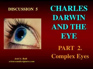
CHARLES DARWIN AND THE EYE DISCUSSION 5 PART 2. Complex Eyes Courtesy Corel Ariel A. Roth sciencesandscriptures.com
OUTLINE1. Complexity of eyes: Special and interdependent parts2. Evolution’s incomplete eye3. The inverted retina: It works very well 4. Conclusions5. Review questions
1. COMPLEXITY OF THE EYE The eye is much more complicated than first surmised. The next figure gives a few more details about our eyes that are basically the “simple” single lens vertebrate eye, also known as the “camera eye. In diagram A, note all the special parts to the right associated with the lens, iris and pupil. These parts are composed of many smaller interdependent parts that are necessary for proper function. Hence, they raise the question of how could these gradually evolving parts provide any evolutionary survival value before all the necessary ones were present so the system could work.
VERTEBRATE EYE A. The complex vertebrate eye. B, C, D, enlarged details.
1. COMPLEXITY OF THE EYE In the slide above note that there are three main coats (layers) to the wall of the eye (diagram C): The tough outer whitish sclera; the middle choroid, that is rich in blood vessels; and the complicated inner retina that is nearly transparent. We will discuss these later when considering the inverted retina. The retina harbors many nerve cells and also the light-detecting cells (photoreceptors) known as the rods and cones (diagram D). The rods function in detecting dim light while the cones detect bright and colored light.
1. COMPLEXITY OF THE EYEDETECTION OF LIGHT One rod may contain 40,000,000 protein molecules called rhodopsin. When light strikes a rhodopsin molecule it bends it. That response is passed on to many more of several different kinds of molecules in an avalanche type of chemical reaction that increases the negative electric charge of the outside of the rod or cone. That change in charge sends an impulse to other nerve cells. In the rod or cone the whole process is reversed in preparation to receive more light. At least a dozen different kinds of protein molecules are involved. Strangely, the eye of the scallop (Pecten) has a double retina, and the inside retina cells become electrically more negative when stimulated - as is the case for man - while in the outside (deeper) retina the cells become more positive. This all adds to the picture we see of a great variety of complex eyes in animals.
1. COMPLEXITY OF THE EYETHE TRILOBITE EYE You may recall that earlier, in the first discussion about Charles Darwin and the eye (No. 4), we mentioned Darwin’s concern about complicated eyes, including their ability to correct for spherical aberration. Spherical aberration prevents a sharp image because parallel light rays coming into the eye do not converge on the same plane. See the two red arrows in the next slide. The even curvature of an ordinary lens is such that light coming in around the outside of the lens focuses on a different plane than light coming through the center, so the image is blurred. There are several ways to correct for spherical aberration. Some trilobites do it by using a very special kind of lens.
ILLUSTRATION OF SPHERICAL ABERRATION. The light rays going through different parts of the lens do not converge (red arrows) on the same plane (retina).
1. COMPLEXITY OF THE EYETHE TRILOBITE EYE The trilobite eye is of special interest because it appears to be one of evolution’s first image-forming eyes that we find in the fossil record as we go up through the geologic layers. We find trilobites in the Cambrian, which is at the bottom of the fossil-rich Phanerozoic part of the geologic column. The next slide is a photograph of Mount Stevens in the Canadian Rockies. The darker layers on the hillside are deep Cambrian rocks that have been obviously pushed up. They contain an abundance of trilobites. The slide following the next is a sample of one of these trilobites from Mount Stevens. The arrow points to the eye region. Note the Canadian coin for scale.
MOUNT STEVENS in the Canadian Rockies. Trilobite fossils are found in the dark middle layers.
CAMBRIAN TRILOBITE FOSSIL. From Mount Stevens in the Canadian Rockies. The red arrow points to the compound eye. Note coin for scale.
1. COMPLEXITY OF THE EYETHE TRILOBITE EYE The trilobite eye, like the eye of an insect, is a compound eye. It has many ommatidia (tubes) each pointing in a slightly different direction, and each ommatidium has its own lens so as to give a precise image of what lies in the exact direction it is pointing. A general diagram of the compound eye used earlier is provided in the next slide for review.
THE COMPOUND EYE. Each ommatidium points in a slightly different direction and detects what is in that direction.
1. COMPLEXITY OF THE EYETHE TRILOBITE EYE In order to get around the problem of spherical aberration, researchers in Europe, such as Descartes and Huygens, working several centuries ago, designed special lenses that corrected for spherical aberration. An example is provided in the next slide. Note that the incoming light rays coming from the right side all converge on the same plane. Amazingly, when the eyes of some trilobites were closely examined it was discovered that their lenses were of the same type as those invented by Descartes. These lenses corrected for spherical aberration and thus provided the trilobite with a sharp image of what it was looking at.
PATTERN OF LIGHT RAYS THROUGH AN APLANATIC LENS. Note the special shape of the lens, and the light rays that converge on one plane (arrow).
1. COMPLEXITY OF THE EYETHE TRILOBITE EYE This sophisticated feat of optical function found in trilobite lenses poses problems for evolution because we don’t find in the fossil record the evolutionary ancestors of these advanced eyes. As evolution would proceed by random mutations, trying one kind of lens shape after another, the number of ineffective shapes tried would be enormous. Yet none have been found.
1. COMPLEXITY OF THE EYETHE TRILOBITE EYE Furthermore, the lenses of trilobites are made of crystals of the mineral calcite (calcium carbonate, CaCO3). Calcite is a complicated mineral that bends the light rays entering or leaving it (index of refraction) at different angles (degree of bending) depending on the orientation of the crystal. In trilobite eyes the calcite of the lenses is oriented in just the proper direction so as to give the right focus. Thus one can wonder about how many random tries it would take before evolution would have produced calcite minerals in the right orientation. And we haven't found the fossils expected for this extended evolutionary process. In several ways the trilobite eye strongly favors the creation concept.
1. COMPLEXITY OF THE EYEA COMMON GENE FOR THE EYE Evolutionist have perceived some evidence for their theory of eye development from a common ancestor based on the genetic makeup of various organisms. It has been found that there is one of those master genes (i.e. Pax 6, a homeobox gene found in many animals) that is associated with the development of the eye in different animals. Evolutionists assume that a common master gene means common evolutionary ancestry. Some complicated genetic engineering experiments conducted in Switzerland have succeeded in taking this eye-inducing gene from a mouse, which has a simple eye, and putting it in the DNA of a fruit fly, which has a compound eye, and that gene caused the development of an extra compound eye on the leg of the fly.
1. COMPLEXITY OF THE EYEA COMMON GENE FOR THE EYE An illustration of this extra eye is shown on the next slide. The eye is to the left of the brown leg. Each of the many bumps on the surface of the white eye is the end of an ommatidium of this compound eye. The ommatidia of this extra eye responded to light by generating a nerve impulse when stimulated by light. So at least the ommatidia were functional.
EXTRA EYE ON THE LEG OF A FLY. Each of the white bumps is an ommatidium of this compound eye.
1. COMPLEXITY OF THE EYEA COMMON GENE FOR THE EYE Evolutionists consider the action of this master gene that causes eye development in different kinds of animals to be strong evidence of a common evolutionary origin. But this needs to be the case only if you assume evolution. On the creation side, it could also mean that one Designer had planned the same kind of basic developmental process in various animals. Why not use the same system of master genes that work in different animals, instead of inventing a different system for each kind of animal? This would seem like efficient planning.
1. COMPLEXITY OF THE EYEA COMMON GENE FOR THE EYE Evolutionists also need to keep in mind that several thousand genes are involved in the development of the eye of the fly and that eye is very different from that of a mouse eye. Evolution needs to account for all these new genes. So one similar master gene does not at all solve the problem of the great variety of different genes producing the variety of eyes that we find.
1. COMPLEXITY OF THE EYETHE SCANNING EYE OF COPILIA We mentioned earlier (Discussion 4) the intriguing eye system of the copepod Copilia. Recall that the animal lives in the Mediterranean Sea and is only about one millimeter wide, yet it uses a scanning system that goes back and forth to form an image, somewhat like a television camera does. The system is illustrated in the next slide. The animal uses four lenses, two in front for viewing and two behind to scan the image captured by the viewing lenses. Muscles cause the scanning lenses (green arrow) to vibrate back and forth about once per second or faster as it views the image seen by the viewing lenses (red arrow).
THE SCANNING SYSTEM. An image is formed by a vibrating scanning lens (green arrow) analyzing the image brought into focus by a viewing lens (red arrow).
1. COMPLEXITY OF THE EYETHE SCANNING EYE OF COPILIA Such an eye provides another example of the great variety of different basic kinds of eyes we find. It does not seem possible that these very different visual systems would evolve from each other. The Copilia eye also illustrates the difficulty of evolving complex systems. For instance in evolving this kind of eye, of what use would be the muscle that vibrates the scanning lens without the evolution of the scanning lens, and of what use would the scanning lens be without a special complicated system in the brain to interpret the scans? Here, as usual, there seem to be too many interdependent parts that are necessary to provide survival value until all are present. Random mutations would not be expected to suddenly provide all the parts of complex working systems so that there could be some survival value.
1.COMPLEXITY OF THE EYETHE TROCHLEA There is a simple ring-like structure associated with our eyes that raises the same kind of question as Copilia does. How could such interdependent parts ever evolve gradually by an unguided random evolutionary process? The structure, called the trochlea, is illustrated at the end of the red arrow in the next slide. A tendon that pulls the eye up and forward slides through that ring so as to change the direction of motion provided by the superior oblique muscle.
ARRANGEMENT OF THE SUPERIOR OBLIQUE EYE MUSCLE. The tendon (tan) of the muscle passes through the ring-like trochlea (red).
1.COMPLEXITY OF THE EYETHE TROCHLEA In an evolutionary process of modification, that needs to provide advantageous survival value in order to succeed, one can wonder how these interdependent parts ever gradually evolved? Did the trochlea ring evolve first? It would be a useless encumbrance by itself. Did the tendon become longer first, so it could extend through the trochlea? Its excess length would negate the usefulness of the muscle. Or did the mechanism that threads the tendon through the trochlea evolve first? That would be useless without first having both a long tendon and a trochlea. You need at least all three factors at the same time to provide evolutionary survival value. Interdependent parts pose serious challenges to evolution.
1. COMPLEXITY OF THE EYE OTHER EXAMPLES OF INTERDEPENDENT PARTS: (a) The brain system that adjusts the focus of the lens is useless without special muscles that change the shape of the lens and a mechanism that determines that the eye is out of focus. (b) The mechanism that adjusts the size of the pupil is useless without a mechanism that detects how much light is present. (c) An eye is useless without a part of abrainto interpret what is seen. (d) Many specialized protein molecules are dependent on each other in order to produce the complex molecular avalanche light detection system mentioned above.
1. COMPLEXITY OF THE EYE OTHER EXAMPLES OF INTERDEPENDENT PARTS: On the next slide is a picture of an eye. While it looks quite simple, behind what you see are the intricate systems mentioned above. Recall that there is no survival value to parts of systems that don’t work unless other necessary parts are also present.
1. COMPLEXITY OF THE EYECONCLUSIONS1. The very complex eyes of trilobites with sophisticated optics appear very early in the fossil record of animals. How could such complexity gradually evolve without leaving any fossil record?The abrupt appearance of such complex functions is better explained by creation.2.The major problem with the evolution of the eye is generally ignored by evolutionists. Complex systems with interdependent parts like the visual system of Copilia, have no evolutionary survival value until all essential parts are present so as to be able to provide the needed survival value. Until then, excess non-functioning parts are only cumbersome impediments. This is the irreducible complexity problem.
1. COMPLEXITY OF THE EYECONCLUSIONSCharles Darwin, in 1859, in his famous book, Origin of Species, (p 219, Penguin Edition, 1968) obviously did not understand the problem of interdependent parts. He states:“If it could be demonstrated that any complex organ existed, which could not possibly have been formed by numerous, successive, slight modifications, my theory would absolutely break down. But I can find out no such case.”Darwin tries to protect his view by using the “just prove it is impossible” type of argument when he says “not possibly.” But his “numerous, successive, slight modifications” that would not have any survival value until something worked, indicates that, in his own words, his theory has “absolutely” broken down many times.
2.EVOLUTION’S INCOMPLETE EYESome evolutionists in Sweden have tried to suggest that the eye could evolve very fast. The reference for their study is:Dan-E Nilsson, Susanne Pelger(Lund University) 1994. A Pessimistic Estimate of the time required for an eye to evolve. Proceedings Royal Society of London, B 256:53-58.These authors conclude that the eye could have evolved in just 1829 steps of arbitrary 1% theoretical improvements.Furthermore, they suggest that it would take less than 364,000 years for a simple eye to evolve from a light sensitive patch.They conclude that there is enough geologic time since the Cambrian for “eyes to evolve more than 1500 times.”The main steps in their proposed model are illustrated in the next picture.
EVOLUTIONOF THE EYE. Cross sections of four stages in gradual development. After Nilson & Pelger, PRSL B 256:53-58.
2.EVOLUTION’S INCOMPLETE EYETheir proposed model cannot be taken seriously because many important parts of the eye are not considered. Their approach is reminiscent of what is sometimes called “fact-free science.” While their valiant efforts are worthy of some respect, the argumentation illustrates the all too common great weakness of many evolutionary propositions, namely, details are overlooked. MISSING PARTS:1. Retina (the most important and most complex part of the eye) 2. Brain parts to interpret what the eye sees3. Nerve connection between eye and brain4. Lens focusing mechanism5. Pupil size adjusting mechanism6. A functional lens (they make a vague suggestion)7. New embryological process needed for vertebrates, where the eye originates from the brain, not the skin as they propose8. Muscles that move the eye
2.EVOLUTION’S INCOMPLETE EYEDespite these major omissions, some evolutionists were excited about the model. Some of their endorsing comments follow:Richard Dawkins,Oxford University. 1994. The Eye in a Twinkling. Nature 368:690-691. Results were “swift and decisive” and the time required for the evolution of the “eye is a geological blink.”Daniel Osorio,Sussex University. 1994. Eye evolution: Darwin’s Shudder Stilled.Trends in Ecology & Evolution 9:241-242. The eye has been such a problem for evolution that it is sometimes referred to as “Darwin’s shudder”INTERNET:“The eye has turned out to be the BEST PROOF of evolution.” (This overstatement has since been removed from its original web page!) To a significant degree these comments, that are highly inaccurate, likely reflect over-reactions by evolutionists to the problem the eye has posed for them for over two centuries.
3. THE INVERTED RETINA Many evolutionists claim that the eye is badly designed! They claim that the retina of vertebrates (fish, amphibians, reptiles, birds and mammals) is inverted from the way it should be. In most other animals they consider it to be verted or properly arranged. The claim of inversion is based on the fact that in vertebrates the light sensitive part (discs) of the photoreceptor cells (rods and cones) is turned away from the light instead of towards it. This is analogous to turning a surveillance camera towards a wall instead of into an open room.
3. THE INVERTED RETINA The next slide illustrates the cells in the two main kinds of arrangements for the retina. Note the direction the light travels, and note the location of the light sensitive area of the photoreceptors (yellow color). The upper figure is the verted arrangement as found in squids, spiders and many snails, etc. The lower figure is the inverted arrangement as found in vertebrates like you. Here, not only does the light have to first go through part of the rod and cone cells to reach the light sensitive discs, it has to also go through layers of neurons; and many evolutionists consider this to be a very bad design.
TWO KINDS OF RETINAS. Note the light sensitive area towards the light in the verted retina and away from the light in the inverted retina.
3. THE INVERTED RETINA In the next slide, used earlier in discussing convergence, you have examples of eyes with these two kinds of retinas. The left is that of a squid that is verted, and the right is that of a vertebrate that is inverted. At this scale, you can barely notice the difference within the thin retinas.
3. THE INVERTED RETINA The two kinds of retinas are quite different in microscopic details. The cephalopod retina has numerous elongated microvilli (illustrated three slides back) that contain the light sensitive molecules, while in vertebrates those light sensitive molecules are found in discs that are constantly being replaced. In the next slide, there are some details of the inverted vertebrate retina. Note especially parts C and the discs in part D. Light comes in from the right.
VERTEBRATE EYE A. The complex vertebrate eye. B, C, D, enlarged details.
3.THE INVERTED RETINADEROGATORY COMMENTS BY SOME EVOLUTIONISTSGeorge Williams.NY University, Stony Brook“There would be no blind spot if the vertebrate eye were really intelligently designed.”Jared Diamond.University of California at Los Angeles“However the vessels and nerves aren’t located behind the photoreceptors, where any sensible engineer would have placed them, but out in front of them, where they screen some of the incoming light. A camera designer who committed such a blunder would be fired immediately. … By contrast, the eyes of the lowly squid, with the nerves artfully hidden behind the photoreceptors are an example of design perfection. If the Creator had indeed lavished his best design on the creature he shaped in his own image, creationists would surely have to conclude that God is really a squid.”
3.THE INVERTED RETINADEROGATORY COMMENTSDouglas Futuyma.University of Michigan and NYUSB“The human eye has a ‘blind spot,’…. It is caused by the functionally nonsensical arrangement of the axons of the retinal cells which run forward into the eye.”William Thwaites.San Diego State University“Vertebrates are cursed with an inside-out retina in the eye…. Did God at the time of the ‘Fall’ turn the vertebrate retina inside-out…?”Richard Dawkins.Oxford University“Any engineer…. would laugh at any suggestion that the photocells might point away from the light, with their wires departing on the side nearest to the light,…. Each photocell is, in effect, wired backwards.”
3.THE INVERTED RETINADEROGATORY COMMENTSThe conclusion is that the eye is so badly designed, that no intelligent designer would commit such a blunder. The implication is that there is no God.THE ALLEGED PROBLEMS OF THE REVERSED OR INVERTED RETINA ARE:a. The light sensitive ends (discs) of the rods and cones are directed (headed) away from the light.b. The nerve processing cells in the retina are between the incoming light and the rods and cones.c. This necessitates a blind spot where the nerve fibers leave the eye to connect to the brain. You may be able to note all these assumed problems on the image of the vertebrate eye repeated in the next slide. The blind spot is labeled “Optic disc” on part A.