Download
1 / 22
220 likes | 907 Views
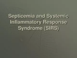
Outline. Definitions and Diagnostic CriteriaPathophysiologyPrognosisTreatmentExample Cases. What is SIRS?. The systemic inflammatory response syndrome is systemic level of acute inflammation, that may or may not be due to infection, and is generally manifested as a combination of vital sign abnormalities including fever or hypothermia, tachycardia, and tachypnea..

E N D
1. Sepsis and the Systemic Inflammatory Response Syndrome
2. Outline Definitions and Diagnostic Criteria
Pathophysiology
Prognosis
Treatment
Example Cases
3. What is SIRS? The systemic inflammatory response syndrome is systemic level of acute inflammation, that may or may not be due to infection, and is generally manifested as a combination of vital sign abnormalities including fever or hypothermia, tachycardia, and tachypnea.
4. Definitions Severe SIRS � SIRS in which at least 1 major organ system has failed.
Sepsis � SIRS which is secondary to infection.
Severe Sepsis � Severe SIRS which is secondary to infection.
Septic Shock � Severe sepsis resulting in hypotensive cardiovascular failure.
5. Criteria for SIRS Requires 2 of the following 4 features to be present:
Temp >38.3� or <36.0� C
Tachypnea (RR>20 or MV>10L)
Tachycardia (HR>90, in the absence of intrinsic heart disease)
WBC > 10,000/mm3 or <4,000/mm3 or
>10% band forms on differential
6. Criteria for Severe SIRS Must meet criteria for SIRS, plus 1 of the following:
Altered mental status
SBP<90mmHg or fall of >40mmHg from baseline
Impaired gas exchange (PaO2/FiO2 ratio<200-250)
Metabolic acidosis (pH<7.30 & lactate > 1.5 x upper limit of normal)
Oliguria (<0.5mL/kg/hr) or renal failure
Hyperbilirubinemia
Coagulopathy (platelets < 80,000-100,000/mm3, INR >2.0, PTT >1.5 x control, or elevated fibrin degredation products)
7. Relationship Between SIRS and Sepsis
8. Risk Factors for SIRS/Sepsis Extremes of age
Indwelling lines/catheters
Immunocompromised states
Malnutrition
Alcoholism
Malignancy
Diabetes
Cirrhosis
Male sex
Genetic predisposition?
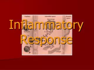
9. Pathophysiology
10. Pathophysiology Vasodilation:
Activation of ATP-sensitive K+ channels in the vascular smooth muscle
Increased synthesis of NO as a result of increased levels of the enzyme, inducible NO synthase
Deficiency of vasopressin
Intravascular Volume Depletion:
Increased capillary permeability leading to third-spacing of fluid
Concurrent volume loss from vomiting or diarrhea
11. Prognosis Overall mortality from SIRS/sepsis in the U.S. is approximately 20%. Mortality is roughly linearly related to the number of organ failures, with each additional organ failure raising the mortality rate by 15%.
Hypothermia is one of the worst prognostic signs. Patients presenting with SIRS and hypothermia have an overall mortality of ~80%.
12. Treatment Fluid Resuscitation
Vasopressors
Antibiotics
Eradication of infection
Ventilatory support, activated protein C, steroids, glycemic control, nutrition
13. Treatment(Fluid Resuscitation) Rapid, large volume infusions are generally indicated in all patients with septic shock.
Some patients require up to 10L of crystalloid in the first 24 hours, with an average requirement of 4-6L.
Although resuscitation with colloid will necessitate less overall volume of fluid, there is no difference between patients treated with colloid versus crystalloid in the development of pulmonary edema, length of stay, or survival.
14. Treatment(Vasopressors) These are second line agents in the treatment of septic shock (after volume resuscitation).
A goal MAP should be 60-65mmHg, although urine output, mental status, and skin perfusion are better variables to use in monitoring adequate perfusion.
15. Treatment(Antibiotics) Empiric antibiotic therapy should be instituted immediately after appropriate cultures have been drawn, taking into consideration the likely source of infection,
In general, therapy should include two effective agents from different classes, for example, a beta-lactam and an aminoglycoside
16. Treatment(Mechanical Ventilation) Nearly all patients with septic shock require supplemental oxygen, and approximately 80% require mechanical ventilation.
Use of mechanical ventilation not only may improve oxygenation, but the necessary sedation +/- paralysis may improve organ perfusion by diverting blood flow away from the diaphragm.
17. Treatment(Activated Protein C) The PROWESS trial showed that patients who received a 96hr infusion of APC within 24 hours of presentation had a statistically lower 28-day mortality rate (25% vs. 31%).
Treatment was of greater benefit in the most acutely ill patients (APACHE II score = 25).
APC has been found to not be cost effective in those patients with APACHE II scores <25 or in those with relatively low life-expectancy even in the event of survival from sespis.
18. Protocol for Early Goal Directed Therapy in Septic Shock
19. Case 1 An 82 year old nursing home resident is brought to the ER by ambulance for an intermittent fever that has lasted about 36 hours. She is currently complaining of some chills and a sensation of thirst. Her past medical history is significant for diabetes, hypertension, and COPD. Her vitals on exam: T=38.6, HR=95, BP=128/65, RR=16, O2 sat=94% on RA. She is mildly diaphoretic, but the remainder of her exam is otherwise normal.
20. Case 2 A 45 year old homeless IV drug user is brought to the ER by a friend because �he looked really sick today and wasn�t making any sense�. He has had recent admissions to the hospital for alcohol withdrawal, and is known to have early cirrhosis without a history of encephalopathy. His vitals on presentation: T=39.5, HR=137, BP=114/40, RR=18, O2 sat=96% on RA. On exam, he appears both acutely and chronically ill, and is lethargic and only oriented to self. He has crackles at the right lung base, a 2/6 systolic murmur at the right upper sternal border. His abdomen is benign, however he has a severe cellulitis with underlying fluctuance beneath his right forearm.
21. Case 3 As the on-call intern, you are called to attend to a cross-cover patient on a non-monitored bed � a 56 year old alcoholic admitted yesterday for alcoholic pancreatitis. Upon admission, he was placed on D5 �NS at 125cc/hr, made NPO, and given morphine prn for pain control. During sign-out, you were told he had �mild pancreatitis� and that there �is nothing to do�. However, when the nursing assistant went in to take his vitals, she found him to be in mild to moderate respiratory distress and �looking bad�. His vitals at this time: T=36.5, HR=140, BP=125/50, RR=34, O2 sat=85% on 4L. Upon exam, he is tremulous, but alert and oriented. His lungs have diffuse crackles bilaterally. His abdomen is moderately tender in the epigastric region, but without peritoneal signs. The patient is currently complaining of shortness of breath and states that he feels �terrible�.
22. Case 4 A 67 year old man with metastatic colon cancer is brought to the ER by his wife because he developed a �high fever� and was �just not looking very well� for the past day. She became stuck in traffic on the way to the hospital, and during the 60 minute trip he has become confused and �sweaty�. His vitals upon arrival: T=40.5, HR=135, BP=76/30, RR=34, O2 sat=84% on RA. On exam, he appears cachectic and acutely ill. He is lethargic and is mumbling incoherently. His lung exam is significant for moderate bilateral crackles. Cardiac exam is significant only for a regular tachycardia. His abdomen appears mildly tender diffusely, but without peritoneal signs. The remainder of the exam is unremarkable.