Download
1 / 56
560 likes | 600 Views
Learn about the anatomy of the female reproductive system, including the external genital organs (vulva) and internal genital organs (vagina, cervix, uterus, fallopian tubes, and ovaries), their functions, and important aspects of each structure.

E N D
Reproductive Health NursingNUR 324Lecture 1Part 1+2Anatomy of the Female Reproductive System
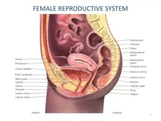
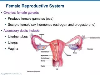
Anatomy of female reproductive system consists of: • Bony pelvis & soft tissue soft tissue consists of: • External female genital organs {The Vulva}. • Internal female genital organs.
External female genital organs {The Vulva} consists of the following structures • The mons pubis: is a pad of fat, covered with pubic hair from the time of puberty Function: protection of the symphysis pubis during intercourse Location: over the symphysis pubis.
The Vulva consists of the following structures 2. The labia majora: are two folds of fat and areolar tissue, covered with skin and pubic hair on the outer surface. Function: protection of the vaginal introitus Location: arise in the mons pubis and merge into the perineum behind.
The Vulva consists of the following structures 3. The labia minora : two thin folds of skin lying between the labia majora. Anteriorly, divide to enclose the clitoris, posteriorly, fuse forming the fourchette. Function: is erotic, in response to stimulation and are highly sensitive. Location: between the labia majora
The Vulva consists of the following structures 4. The clitoris: (corresponding to the male penis). is a small rudimentary organ, very sensitive and highly vasculars • Function: is sexual stimulation.( plays a part in the orgasm of sexual intercourse.)
The Vulva consists of the following structures 5. The vestibule: Location: the area in which the openings of the urethra and the vagina are situated & enclosed by the labia minora. 6. The urethral orifice Location :2.5 cm posterior to the clitoris
The Vulva consists of the following structures 7. The Vaginal orifice (Vaginal introitus) Location :occupies the posterior two-thirds of the vestibule. The orifice is partially closed by the hymen 8. The Hymen, a thin membrane which tears during sexual intercourse . It has one or more openings to allow escape of menstrual blood.
The Vulva consists of the following structures 9. Bartholin's glands: are two small glands which open on either side of the vaginal orifice Function: secrete mucus, which lubricates the vaginal opening. Location: in the posterior part of the labia majora
INTERNAL GENITALIA The internal genitalia consists of the: • Vagina • Cervix • Uterus • Fallopian Tubes • Ovaries
VAGINA • The vagina connects the cervix to the external genitals • It is located between the bladder and rectum • It functions : -As a passageway for the menstrual flow -For uterine secretions to pass down through the introitus -As the birth canal during labor • Opening may be covered by a thin sheath called the hymen
PERINEUM • The muscle and tissue located between the vaginal opening and anal canal • It supports and surrounds the lower parts of the urinary system • The perinium contains an abundance of nerve endings that make it sensitive
CERVIX • The cervix connects the uterus to the vagina • The cervical opening to the vagina is small • This acts as a safety precaution against foreign bodies entering the uterus • During childbirth, the cervix dilates to accommodate the passage of the fetus • This dilation is a sign that labor has begun
UTERUS • Commonly referred to as the womb • A pear shaped organ • The powerful muscles of the uterus expand to accommodate a growing fetus and push it through the birth canal
UTERUS Layers of the Uterine Wall • Outer layer = the Serosa or perimetrium • Middle layer = the muscularis or myometrium. • The Inner layer = the mucosa of the endometrium.
UTERUS Function of uterus: shelter the fetus during pregnancy. It prepares for this possibility each month and following pregnancy it expels the uterine contents. Location: situated in the true pelvis , between the bladder and rectum.( In pelvic cavity)
FALLOPIAN TUBES • Serve as a pathway for the ovum to the uterus • Are the site of fertilization by the male sperm • Often referred to as the oviducts or uterine tubes • Fertilized egg takes approximately 6 to 10 days to travel through the fallopian tube to implant in the uterine lining
FALLOPIAN TUBES Functions: 1-Receives the spermatozoa as they travel upwards 2-Ovum transport and pick up. 3- provides a site for fertilization. 4-Embryo transport and nourishment.
OVARIES 1. The ovaries: (female gonads) comparable to the testes in the male & similar to almonds in size & shape. Location: Are located on either side of the uterus ,below and behind the fimbriated ends of the ova ducts. Functions: The ovaries produce ova (ovulation) and the hormones estrogen, progesterone.
OVARIES • The female glands • They develop and expel an ovum each month • A woman is born with approximately 400,000 immature eggs called follicles • During a lifetime a woman release about 400 to 500 fully matured eggs for fertilization • The follicles in the ovaries produce the female sex hormones, progesterone and estrogen • These hormones prepare the uterus for implantation of the fertilized egg
BREASTS Breasts may exhibit cyclical changes, including increased swelling and tenderness prior to menstruation
Placenta Functions of the Placenta • Transfers blood and nutrition from the mother to the fetus. • Transfer waste product from the fetal metabolism to the maternal circulation for disposal. • Maternal circulation is separated from the fetal circulation The umbilical cord consist of two arteries and one vein
Follicle stimulating hormone FSH Luteinizing hormone LH-signals ovulation Estrogen- produced throughout the menstrual cycle Progesterone-produced during second half of cycle Both FSH and LH are produced in the pituitary gland Both estrogen and progesterone are produced by the follicles in the ovaries SEX HORMONES
Menarch, the onset of menstruation signals the bodily changes that transform a female body Average age is 12yrs Amount of bleeding varies from woman to woman Expulsion of blood clots Blood color can vary from bright red to dark Usually occurs every 25 to 32 days Women can experience fluid retention, cramping, mood swings, weight gain, breast tenderness, diarrhea/ constipation MENSTRUATION
Biology and the Menstrual Cycle • Menstrual cycle is regulated by fluctuating levels of sex hormones. • These hormones produce certain changes in the ovaries and uterus.
The Phases of the Menstrual Cycle • 1. Follicular phase • 2. Ovulation • 3. Luteal phase • 4. Menstruation
What Happens in the OvariesDuring the Menstrual Cycle • Follicular phase - high levels of FSH secreted. • Function is to stimulate follicles in the ovaries. • One follicle begins to ripen and brings an egg to maturity. • Follicle secretes estrogen. • Ovulation - follicle ruptures open and releases the ripened egg.
What Happens in the Ovaries During the Menstrual Cycle • Luteal phase - after releasing an egg, the follicle turns into the corpus luteum andmanufactures progesterone. • Menstruation - shedding of the inner lining of the uterus.
What Happens in the Uterus During the Menstrual Cycle • Follicular phase - high levels of estrogen stimulate the endometrium. • Luteal phase - progesterone secreted by the corpus luteum stimulates the glands of the endometrium to start secreting nourishing substances.
What Happens in the Uterus During the Menstrual Cycle • Corpus luteum continues to produce estrogen and progesterone for about 10 to 12 days. • If pregnancy has not occurred, hormone output declines. • Menstrual fluid is combination of blood from the endometrium, degenerated cells, and mucus from the cervix and vagina. • The ovaries call the shots in regulating the cycle.
Length and Timing of the Menstrual Cycle • Normal menstrual cycle = 20 to 40 days; average is about 28 days. In an average cycle: • Menstruation begins on day 1 and continues until about day 4 or 5. • Follicular phase - about days 5-13. • Ovulation occurs on day 14. • Luteal phase - day 15 to the end of the cycle, day 28.
Conception • Conception is the union of an egg from the mother and sperm from the father – these are germ cells or gametes • Gametes contain only half of the genetic material (23 chromosomes) found in other cells
Fertilization • Starts with the release of an egg from the woman’s ovaries • Sperm swim up the female reproductive tract for about 6 hours aided by muscular contractions of the uterus • From millions that enter the vagina only about 200 get near the egg
Journey of Ovum Zygote Ovum Mature ovum in follicle
When the first sperm enters, the egg changes in the zona to prevent entry of other sperm. • The nuclei of ovum and sperm unite to form a zygote.
Fertilized Ovum 2 individual cells
From Zygote to Embryo • Cell division The zygote divides into 2, 4, eight, etc. A hollow ball of cell forms called the blastocyst • Cell migration Cells move to go somewhere else and form 3 different layers • Cell differentiation Cells take on specific structure and function.