Download
1 / 1
10 likes | 148 Views
Hyaluronan oligosaccharide based biomaterials for peripheral nerve regeneration Kathryn A. Bivens 1 , Christine E. Schmidt 2 The University of Texas at Austin 1 Department of Chemical Engineering 2 Department of Biomedical Engineering.

E N D
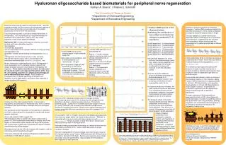
Hyaluronan oligosaccharide based biomaterials for peripheral nerve regeneration Kathryn A. Bivens1, Christine E. Schmidt2 The University of Texas at Austin 1Department of Chemical Engineering 2Department of Biomedical Engineering • Peripheral nerve injuries require an estimated 50,000 – 200,000 surgeries annually, with the common clinical treatment requiring a graft of tissue removed from a secondary site in the patient, causing loss of function from the second site. • Tissue engineering seeks to provide an effective alternative to this approach. Our long-term goal is to engineer therapies for peripheral nerve regeneration, making use of the unique properties of hyaluronan based biomaterials. • Hyaluronan (HA) is a glycosaminoglycan naturally occurring in the body, with many desirable properties: • biocompatible • enzymatically degradable • can be crosslinked to form hydrogel networks of varying porosity and degradation rate • involved in scarless wound healing and angiogenesis (West et al., 985) • HA fragments have biological activity in many cell types • HA and HA receptors play a role in axon-Schwann cell interactions and nerve repair (Sherman et al., 2000; Eggli et al., 1996) • We are interested in understanding the role of HA fragments in nerve regeneration and in ultimately creating materials that release desirable sizes and concentrations of therapeutic HA fragments. These studies describe our efforts to understand the mechanism of the size-dependent bioactivity of HA, how the cell signaling properties of a simply glycosaminoglycan can be modulated by chain length. Future studies will investigate how this applies specifically to Schwann cell regulation in nerve regeneration, and seek to tailor materials to release beneficial HA fragments. • 13Carbon NMR spectra of HA oligosaccharides, • illustrating the contribution of size-related conformational changes to acetamido C=O resonance. • HA fragments of ~2-4 disaccharides • HA fragments of ~12 disaccharides • HA fragments of ~7 disaccharides • HA fragments of ~10 disaccharides • HA fragments of ~27 disaccharides • HA fragments of ~59 disaccharides • HA fragments of ~99 disaccharides • Very small HA fragments (a), at the bottom of the bioactive range, give a high, sharp, internal acetamido C=O peak, associated with hydrogen bonding within the same HA molecule or with the unbonded carbon. • They also show two additional pronounced peaks associated with terminal acetamido C=O. • As oligosaccharides increase in size, end group contributions become imperceptibly small: (d), (e). • As oligosaccharide size increases, the peak representing internal acetamido C=O resonance becomes shorter and broader, indicative of increased intermolecular hydrogen bonding. • Compare (b) and (c) to (f) and (g). • The biological activity of the same oligosaccharide sizes was tested by assessment of their ability to promote human aortic endothelial cell proliferation. After 48 hours in culture, HA fragments of less than 60 disaccharides in length appeared to Analysis of structure transition by measuring changes in 13carbon NMR resonance supports the idea that the structure of HA in solution undergoes a change at around 60 disaccharides in length. This also appears to correlate with the size-dependent effects of HA fragments on endothelial cell proliferation, suggesting that this transition from secondary to tertiary structure may be responsible for the differing bioactivities of different sizes of HA. (a) (b) 13Carbon NMR spectrum of 2-4 disaccharide HA. Peak at ~174 ppm indicates carboxyl resonance, which is not strongly affected by state of hydrogen bonding.Peaks at ~174.4 and 174.8 ppm represent terminal acetamido C=O resonance.Peak at ~175.4 represents internal acetamido C=O resonance, which is strongly affected by state of hydrogen bonding.(Scott and Heatley, 2002; Cowman et al., 1996) • 13Carbon NMR spectra of the carbonyl region of high molecular weight HA. • HA under normal conditions, with a broad acetamido C=O resonance at ~175 ppm. • HA ‘denatured’ at high pH, with acetamido C=O resonance sharpened, heightened, and shifted slightly upfield. This shift is indicative of the disruption of intermolecular hydrogen bonds. • (Scott and Heatley, 2002) (c) CD44 preferentially binds to HA chains as small as 3 disaccharides in length, while chains as small as 4 disaccharides promote angiogenesis(Underhill and Toole, 1979; West et al., 1985). This relatively simple ligand stimulates CD44 receptors through a possible mechanism of receptor clustering, while the same polymer when roughly an order of magnitude larger does not provide a signal. Future studies will attempt to refine our picture of the structural transition of HA in solution, adding 13carbon NMR measurement of oligosaccharides of ~80 and ~200 disaccharides, and repeating measurements of ~27 and ~99 disaccharides to obtain greater resolution. Endothelial cell proliferation experiments will be repeated with longer incubation periods to determine significance. To better understand CD44-mediated size dependent bioactivity of HA, future studies will include stimulation of CD44 on endothelial cells as well as Schwann cells using antibodies, antibody fragments, and crosslinked antibody fragments in an attempt to reproduce the effects of HA fragments of various sizes. (d) (e) HA molecule (a) (b) (f) membrane membrane Structure of HA, indicating possible states of hydrogen bonding. (a) The reducing terminal end of HA, showing internal hydrogen bonding (dotted line) resulting in a helical secondary structure. The terminal acetamido group ( circled) has no available carboxylate receptor to bind to. (b) Tertiary structure of HA, formed by intermolecular hydrogen bonds, resulting in a β sheet-like structure. Vertical arrows indicate hydrogen bonds between antiparallel HA chains, pointing from donor NH to acceptor COO-(Scott and Heatley, 2002) (g) CD44 is one of the main receptors for HA. It is involved in angiogenesis and stimulation of endothelial cells, and it is present on Schwann cells and involved in Schwann cell-axon neuregulin signaling. One hypothesis for the mechanism of HA fragments for activation via CD44 is that binding of HA molecules of a certain size causes receptor clustering. Ghosh and Guidolin (2002) suggest that "HA preparations within a specific size range could provoke a pattern of CD44 clustering and crosslinking on binding, which then triggers an intracellular signal, whereas the larger HA molecules may occupy these multiple CD44 linking sites but prevent receptor crosslinking and a cellular reaction.“ What determines how the HA chain interacts with receptors, and the critical size at which bioactivity changes? It has been proposed that binding and bioactivity is determined by conformation of HA chains in solution. Highly crosslinked α-CD44 Single α-CD44 induce increased proliferation when compared to negative controls, with fragments of ~4 disaccharides causing significantly more proliferation. Fragments ~99 disaccharides in length were indistinguishable from negative controls. HA can exist in “stiff” vs. “flexible” structures, with stiffness associated with high molecular weight chains in a unique tertiary structure. “Stiffness” is associated with chains as small as 60 disaccharides in length (Darke et al., 1975; Mathews and Decker, 1977) Tertiary structures of large HA chains in solution have been proposed as seen in the figure above. Hydrogen bonding associated with secondary vs. tertiary structure can be assessed by its effects on the 13carbon NMR resonances of groups involved in bonding. We have investigated the structural change occurring through a range of HA oligosaccharides, using 13carbon NMR performed with HA dissolved at 10 mg/ml in D20 with 0.15 M NaCl. Spectra were acquired using a Varian Associates Unity 300 spectrometer. Approximately 30,000 repetitions were captured for each spectrum. * membrane membrane * Ultimately we hope to use this knowledge to tailor release of therapeutic HA oligosaccharides from scaffolds used in peripheral nerve regeneration. • Acknowledgements: • National Science Foundation • UT Austin College of Engineering THRUST Program Human aortic endothelial cells (p6) were seeded in 96 well plates at 10,000 cells/cm2 and allowed to adhere for 2 hours before culturing for 48 hours in starvation medium (EBM-2 with 1% FBS) containing 1.5 μg/ml HA of various sizes. Starvation medium was used as a negative control and EGM-2 as a positive control. For each condition, n=8, with the experiment repeated 3 times. Cell proliferation was measured with the colorimetric MTS assay. * indicates significant difference from negative control, p < 0.01.