Download
1 / 49
490 likes | 507 Views
Dive into the structure and function of DNA, its role in heredity, and the fascinating world of molecular biology, including DNA replication, transcription, and translation. Explore the genetic material that shapes all living organisms.

E N D
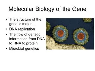
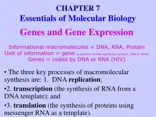
Viruses provided some of the earliest evidence that genes are made of DNA • Molecular biology studies how DNA serves as the molecular basis of heredity
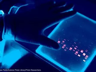
THE STRUCTURE OF THE GENETIC MATERIAL Experiments showed that DNA is the genetic material • The Hershey-Chase experiment showed that certain viruses reprogram host cells to produce more viruses by injecting their DNA Head DNA Tail Tailfiber = Protein coat
Agitate in a blender to separate phages outside the bacteria from the cells and their contents. Centrifuge the mixture so bacteria form a pellet at the bottom of the test tube. Measure the radioactivity in the pellet and liquid. Mix radioactivelylabeled phages with bacteria. The phages infect the bacterial cells. 1 2 3 4 • The Hershey-Chase Experiment Radioactiveprotein Emptyprotein shell Radioactivityin liquid Phage Bacterium PhageDNA DNA Batch 1Radioactiveprotein Centrifuge Pellet RadioactiveDNA Batch 2RadioactiveDNA Centrifuge Radioactivityin pellet Pellet
Phage reproductive cycle – how a virus works Phage attaches to bacterial cell. Phage injects DNA. Phage DNA directs host cell to make more phage DNA and protein parts. New phages assemble. Cell lyses and releases new phages.
DNA and RNA are polymers of nucleotides • DNA is a nucleic acid, a polymer made of long chains of nucleotides • A nucleotide = a phosphate + a 5-carbon sugar + a nitrogenous base Phosphate group Nitrogenous base Nitrogenous base(A, G, C, or T) Sugar Phosphategroup Nucleotide Thymine (T) Sugar(deoxyribose) DNA nucleotide Polynucleotide Sugar-phosphate backbone
DNA has four kinds of bases, A, T, C, and G Thymine (T) Cytosine (C) Adenine (A) Guanine (G) Pyrimidines Purines C5H6N2O2 C5H5N5O C4H5N3O C8H17N
RNA has a slightly different sugar – ribose • RNA has U instead of T • RNA is also a nucleic acid Nitrogenous base(A, G, C, or U) Phosphategroup Uracil (U) Sugar(ribose)
DNA is a double-stranded helix • James Watson and Francis Crick worked out the three-dimensional structure of DNA, based on work by Rosalind Franklin
Abstract from"The Double Helix" by James D. Watson: Abstract from "The Double Helix" by James D. Watson: • ... I quickly cleared away the paper from my desk top so I would have a large, flat surface on which to form pairs of bases held together by hydrogen bonds. Though I went back to my like-with-like prejudices, I saw all to good that they led nowhere....... Suddenly I became aware that an adenine-thymine pair held together by two hydrogen bonds was identical in shape to a guanine-cytosine base pair held together by at least two hydrogen bonds. All the hydrogen bonds seems to form naturally; no fudging was required to make the 2 type of base pairs identical in shape...
The structure of DNA consists of two polynucleotide strands wrapped around each other in a double helix 1 chocolate coat, Blind (PRA) Twist
Each base pairs with a complementary partner • A pairs with T • G pairs with C • Hydrogen bonds between bases hold the strands together
Hydrogen bond • Three representations of DNA Ribbon model Partial chemical structure Computer model
DNA REPLICATION DNA replication depends on specific base pairing • In DNA replication, the strands separate • Enzymes use each strand as a template to assemble the new strands A A Nucleotides Parental moleculeof DNA Both parental strands serveas templates Two identical daughtermolecules of DNA
DNA replication: A closer look • DNA replication begins at several sites Parental strand Origin of replication Daughter strand Bubble Two daughter DNA molecules
5 end 3 end P • Each strand of the double helix is oriented in the opposite direction P P P P P P P 3 end 5 end Figure 10.5B
3 DNA polymerasemolecule 5 5 end Daughter strandsynthesizedcontinuously Parental DNA 5 3 Daughter strandsynthesizedin pieces • How DNA daughter strands are synthesized • The daughter strands are identical to the parent molecule 3 P 5 5 P 3 http://www.ncc.gmu.edu/dna/repanim.htm http://www.johnkyrk.com/DNAreplication.html DNA ligase http://www.stolaf.edu/people/giannini/flashanimat/molgenetics/dna-rna2.swf Overall direction of replication Figure 10.5C
THE FLOW OF GENETIC INFORMATION FROM DNA TO RNA TO PROTEIN The DNA genotype is expressed as proteins, which provide the molecular basis for phenotypic traits • The information constituting an organism’s genotype is carried in its sequence of bases
The DNA is transcribed into RNA, which is translated into the polypeptide • A specific gene specifies a polypeptide DNA TRANSCRIPTION mRNA TRANSLATION Protein Figure 10.6A
Gene 1 Gene 3 DNA molecule Gene 2 DNA strand TRANSCRIPTION RNA Codon TRANSLATION Polypeptide Amino acid Figure 10.7
Genetic information written in codons is translated into amino acid sequences • The “words” of the DNA “language” are triplets of bases called codons • The codons in a gene specify the amino acid sequence of a polypeptide
The genetic code is the Rosetta stone of life • Virtually all organisms share the same genetic code • Read this chart from the mRNA strand to find out the sequence of amino acids Figure 10.8A
About the DNA code…. There are a number of important features of the genetic code outlined below: • The code is a triplet code, i.e. three nucleotides specifies an amino acid in a protein • The code is read continuously from the first methionine start codon to the stop codon at the end. • The RNA is read in successive groups of three nucleotides, without punctuation. The RNA could be read in any of three "frames" but only one is correct (see the 'bla bla bla' overhead). • The code is almost universal, i.e. the same codon encodes the same amino acid in different organisms. There are only a couple of exceptions to this. • The code is degenerate: more than one codon exists for all of the amino acids apart from methionine and tryptophan. • The code has start and stop signals. AUG indicates the start of translation at the start of the RNA molecule (AUG encodes methionine) and sets the correct reading frame at the start of the protein. A number of codons (UAA, UAG, UGA) indicate the end of translation.
How to read the code…. • Translation must start at a defined position in the RNA and the code must be read in codons of 3 bases. • BLABLATHEBOYATETHEBUGBLA • This looks like a load of jibberish in this format. However, if we apply a couple of simple rules it is possible to make sense of this. Here are the 2 rules that we need here: • We only start reading this sentence at the first THE word, equivalent to the first AUG in a mRNA. • We read the sentence only in groups of 3 letters with no punctuation between words. When we apply these rules, this is what we get: • BLABLA THE BOY ATE THE BUG BLA
Transcribed strand An exercise in translating the genetic code DNA Transcription RNA Startcodon Stopcodon Translation Polypeptide Figure 10.8B
Give it a try! DNA Code: ---ATC ATG TTT CCT GAT ACT m-RNA Code: ____ ____ ____ ____ t-RNA Code: -____ ____ ____ ____ Amino Acids: ____ ____ ____ ____
Transcription produces genetic messages in the form of RNA RNA nucleotide RNApolymerase Direction oftranscription Templatestrand of DNA Newly made RNA Figure 10.9A
RNA polymerase DNA of gene Promoter DNA Terminator DNA Initiation • RNA nucleotides line up along one strand of the DNA following the base-pairing rules • The single-stranded messenger RNA peels away and the DNA strands rejoin • In transcription, the DNA helix unzips Elongation Area shownin Figure 10.9A Termination GrowingRNA Completed RNA RNApolymerase Figure 10.9B
Eukaryotic RNA is processed before leaving the nucleus Exon Intron Exon Intron Exon DNA TranscriptionAddition of cap and tail • Noncoding segments called introns are spliced out • A cap and a tail are added to the ends Cap RNAtranscriptwith capand tail Introns removed Tail Exons spliced together mRNA Coding sequence NUCLEUS CYTOPLASM Figure 10.10
Transfer RNA molecules serve as interpreters during translation Amino acid attachment site • In the cytoplasm, the mRNA attaches to a ribosome and translates its message into a polypeptide • The process is aided by transfer RNAs (tRNA) Hydrogen bond RNA polynucleotide chain Anticodon Figure 10.11A
Each tRNA molecule has a triplet anticodon on one end and an amino acid attachment site on the other Amino acidattachment site Anticodon Figure 10.11B, C
Ribosomes build polypeptides Next amino acidto be added topolypeptide Growingpolypeptide tRNA molecules P site A site Growingpolypeptide Largesubunit E site tRNA tRNA P A E mRNA mRNAbindingsite Codons mRNA Smallsubunit Figure 10.12A-C
An initiation codon marks the start of an mRNA message Start of genetic message End Figure 10.13A
mRNA, a specific tRNA, and the ribosome subunits assemble during initiation Largeribosomalsubunit Initiator tRNA P site A site Startcodon Small ribosomalsubunit mRNA 1 2 Figure 10.13B
Elongation adds amino acids to the polypeptide chain until a stop codon terminates translation • The mRNA moves a codon at a time relative to the ribosome • A tRNA pairs with each codon, adding an amino acid to the growing polypeptide
Amino acid ANIMATION: http://www.lewport.wnyric.org/JWANAMAKER/animations/Protein%20Synthesis%20-%20long.html More detailed: http://207.207.4.198/pub/flash/26/transmenu_s.swf Polypeptide Asite P site Anticodon mRNA 1 Codon recognition mRNAmovement Stopcodon Newpeptidebond 2 Peptide bond formation 3 Translocation Figure 10.14
Review: The flow of genetic information in the cell is DNARNAprotein • The sequence of codons in DNA spells out the primary structure of a polypeptide • Polypeptides form proteins that cells and organisms use
TRANSCRIPTION DNA Stage mRNA istranscribed from aDNA template. 1 mRNA RNApolymerase • Summary of transcription and translation Amino acid TRANSLATION Stage Each amino acid attaches to its proper tRNA with the help of a specific enzyme and ATP. 2 Enzyme tRNA Initiator tRNA Anticodon Stage Initiation of polypeptide synthesis 3 Largeribosomalsubunit The mRNA, the first tRNA, and the ribosomal subunits come together. Start Codon Smallribosomalsubunit mRNA Figure 10.15
Newpeptidebondforming Growing polypeptide Stage Elongation 4 A succession of tRNAs add their amino acids to the polypeptide chain as the mRNA is moved through the ribosome, one codon at a time. Codons mRNA Polypeptide Stage Termination 5 The ribosome recognizes a stop codon. The poly-peptide is terminated and released. Stop Codon Figure 10.15 (continued)
Mutations can change the meaning of genes • Mutations are changes in the DNA base sequence • These are caused by errors in DNA replication or by mutagens • The change of a single DNA nucleotide causes sickle-cell disease
NORMAL GENE • Point mutations – error of just one base mRNA Protein Met Lys Phe Gly Ala BASE SUBSTITUTION Met Lys Phe Ser Ala Missing BASE DELETION Met Lys Leu Ala His Figure 10.16B
Normal hemoglobin DNA Mutant hemoglobin DNA mRNA mRNA Normal hemoglobin Sickle-cell hemoglobin Glu Val Figure 10.16A
Single Gene Mutations • Many more human diseases result from single gene mutations than from chromosome abnormalities. • sickle-cell anemia • cystic fibrosis • Huntington disease • phenylketonuria (PKU) • alkaptonuria. • These disorders, referred to as inborn errors of metabolism, cause the absence of, or incorrect form of, the protein for which that gene codes. • Usually autosomal recessive traits. In heterozygotes, the cell's normal gene compensates by making more protein.
Types of Mutations • Missensemutation : change in one base pair, substitution of one amino acid for another in the protein made. • Nonsense mutation: also a change in one base pair, however, the altered DNA prematurely signals cell to stop building a protein. shortened protein that may function improperly or not at all. • Insertion: changes the number of bases in a gene by adding a piece of DNA. protein made by the gene may not function properly. • Deletion: changes the number of bases by removing a piece of DNA. Small deletions may remove one or a few base pairs within a gene, while larger deletions can remove an entire gene or several neighboring genes. may alter the function of the resulting protein(s). • Duplication: a piece of DNA is abnormally copied one or more times. -> may alter the function of the resulting protein. • Frameshift mutation: addition or loss of DNA bases changes a gene’s reading frame (3-letter codons). changes the code for amino acids. Resulting protein is usually nonfunctional. Insertions, deletions, and duplications can all be frameshift mutations. • Repeat expansion: Short DNA sequences are repeated several times in a row normally in DNA. An expansion increases the number of repeats. can cause the resulting protein to function improperly.