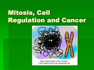
Cell Cycle Regulation and Cancer
Cell Cycle Regulation and Cancer. Lecture #15 Honors Biology Ms. Day. Another Type of Cell Division: Binary Fission. Prokaryotes (bacteria) Reproduce by a type of cell division called binary fission No nucleus no karyokinesis!!. Origin of replication. Cell wall. Plasma Membrane.

Cell Cycle Regulation and Cancer
E N D
Presentation Transcript
Cell Cycle Regulation and Cancer Lecture #15 Honors Biology Ms. Day
Another Type of Cell Division: Binary Fission • Prokaryotes (bacteria) • Reproduce by a type of cell division called binary fission • No nucleus no karyokinesis!!
Origin of replication Cell wall Plasma Membrane E. coli cell Bacterial Chromosome Chromosome replication begins. Soon thereafter, one copy of the origin moves rapidly toward the other end of the cell. Two copies of origin 1 Replication continues. One copy ofthe origin is now at each end of the cell. 2 Origin Origin Replication finishes. The plasma membrane grows inward, and new cell wall is deposited. 3 4 Two daughter cells result. • In binary fission, • The bacterial chromosome replicates • The two daughter chromosomes move apart Figure 12.11
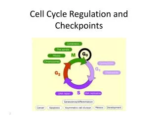
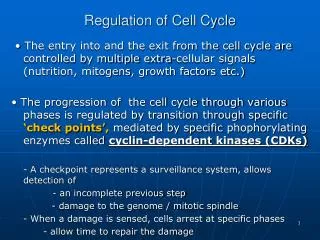
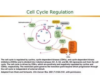
Cell Cycle needs to be controlled (Regulated) • The cell cycle has 3 checkpoints • A place where stop and go signals can regulate (control) cycle • Signals make sure things • been completed • completed correctly • There are 3 checkpoints • G1 checkpoint • G2 Checkpoint • M checkpoint
G1 checkpoint Control system S G1 G2 M M checkpoint Figure 12.14 G2 checkpoint The Cell Cycle Control System
G0 G1 checkpoint G1 G1 If a cell does not receive a go-ahead signal at the G1checkpoint, cell exits the cell cycle and goes into G0, a nondividing state. If a cell receives a go-ahead signal at the G1 checkpoint, the cell continues on in cell cycle. Figure 12.15 A, B Checkpoint = give “go” or “stop” signals
Chromosomes are lined up in the middle properly of replicated DNA of unreplicated (original) DNA http://highered.mcgraw-hill.com/sites/0072495855/student_view0/chapter2/animation__control_of_the_cell_cycle.html
What controls the checkpoints? • Two types of proteins in cytoplasm • Cyclins • cyclin-dependent kinases (Cdks)
INACTIVE FORM CYCLIN DEPENDENT KINASE (CDK) CYCLIN + ACTIVE FORM CDK/CYCLIN COMPLEX
Active vs. Inactive?? • What happens when cyclins and cdks are in the ACTIVE form? • Cells can pass through the cell cycle to the NEXT phase • What happens when cyclins and cdks are in the INACTIVE form? • Cells can NOT pass through the cell cycle to the NEXT phase
cyclin degrades & breaks apart cyclin degrades & breaks apart
What degrades (breaks down) cyclins? • Proteolytic enzymes (proteins) • Break down/degrade cyclins cause them to fluctuate in [ ] • “PROTEO” means protein • “LYTIC” means break or lyse REMEMBER: • Cyclin concentration fluctuates (changes) • Cdk concentration stays the SAME
2 µm Figure 21.17 Programmed Cell Death (Apoptosis) • If cell doesn’t “pass” checkpoint, it goes through apoptosis • http://www.dnatube.com/video/1188/Apoptosis-animation • Cell signaling is involved in programmed cell death needed to maintain healthy tissues/ cell function http://bio-alive.com/categories/apoptosis/apoptosis.htm
What other things control cell division? • Both internal and external signals control the cell cycle/cell division… • Internal signals • CDK/Cyclins at checkpoints • External signals • Growth factors • Density dependent inhibition • Anchorage dependence
External (outside the cell) Influences on Cell Division • Growth factors • Stimulate other cells to divide • In density-dependent inhibition • Crowded cells stop dividing • Most animal cells exhibit anchorage dependence • Cells must be attached to a structure to divide • Ex: protein of a tissue or another cell
(a) Cells anchor to dish surface and divide (anchorage dependence). When cells have formed a complete single layer, they stop dividing (density-dependent inhibition). Normal mammalian cells. **The availability of nutrients, growth factors, and a substratum for attachment limits cell density to a single layer. If some cells are scraped away, the remaining cells divide to fill the gap and then stop (density-dependent inhibition). Figure 12.18 A 25 µm
Loss of Cell Cycle Controls in Cancer Cells • Abnormal dividing cells form tumors • TUMOR= mass or group of abnormal dividing cells
Abnormally Dividing Cells make Tumors • Two Types of Tumors: • Benign “fine” • Clump of cells remain at orginal spot • Malignant “mean” “cancer” • Loose/destroy attachments to other cells they can spread or move (called metastasize)!!!
Cancer cells usually continue to divide well beyond a single layer, forming a clump of overlapping cells. Figure 12.18 B 25 µm Cancer cells = continue to divide w/out rules • Do NOT follow the “rules” • No checkpoints and no density-dependent inhibition or anchorage dependence • Immortal cells (if enough nutrients)
Why do cells break the “rules”? • Don’t need growth factors maybe they make their own growth factors • Mutations in GENES that make proteins involved in control systems!!!
Cancer cells are “hungry”… • Angiogenesis • Making of blood vessels towards tumor • Without blood and the nutrients it carries, a tumor would be unable to continue growing.
Cancer Treatment • Radiation destroys DNA in cancer cells (these cells have lost ability to repair damage) • Chemotherapeutic drugs interfere with specific steps in cell cycle • Also effects normal cells
3 4 1 2 Tumor Lymphvessel Bloodvessel Glandular tissue Cancer cell MetastaticTumor Cancer cells spread through lymph and blood vessels to other parts of the body. A small percentage of cancer cells may survive and establish a new tumor in another part of the body. A tumor grows from a single cancer cell. Cancer cells invade neighboring tissue. Figure 12.19
Movies for Review... Cancer Movie • http://www.cancerquest.org/index.cfm?page=3102&lang=english Angiogenesis • http://www.hhmi.org/biointeractive/angiogenesis