Download
1 / 4
40 likes | 149 Views
Figure 8.16. MRI scans of 10 healthy subjects who saw line draw. ings of faces (red). or buildings (green). The view is from the bottom of the brain. (. Avidan. et al., 2005).

E N D
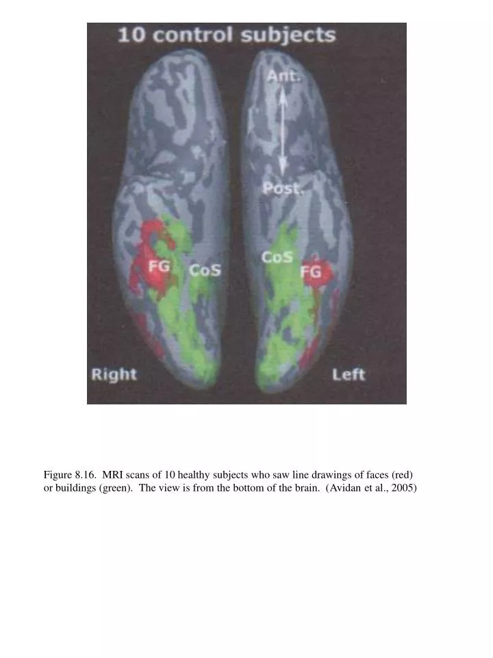
Figure 8.16. MRI scans of 10 healthy subjects who saw line draw ings of faces (red) or buildings (green). The view is from the bottom of the brain. ( Avidan et al., 2005)
Figure 8.17. Activity in 9 human brains in the fusiform face area when faces where implied in the images, even though the faces were presented in a fuzzy manner. The brain activity was scanned using fMRI. (Cox et al., 2004)
a b Figure 8.18. Two paintings by Rene Magritte. a. The Idea. [size, medium, date, location]. b. Not to be reproduced., 1937. Oil on canvas, 79 x 66 cm. Rotterdam, Museum Boymans-van Beuningen.
Figure 8.19. The figure on the left reveals what the prosopagnosic subject looked at to judge whether or not a face was shown. AM stands for age-matched healthy subjects, 54 years old; C stands for healthy young adult subjects. (Caldara et al., 1995).